Latin: musculus sphincter urethrae externus (sphincter urethrae)
Overview
- The voluntary (skeletal-muscle) sphincter of the urethra.
- Part of the muscle layer of the deep perineal pouch (formerly described as the urogenital diaphragm), together with the deep transverse perineal muscle.
- Distance from the external urethral opening: about 1–2 cm in women, about 15–18 cm in adult men (highly variable).
- The internal urethral sphincter, at the neck of the bladder, is smooth muscle and is continuous with the detrusor muscle.
- In women the external sphincter is more complex, with three parts (sphincter urethrae, urethrovaginal sphincter, compressor urethrae).
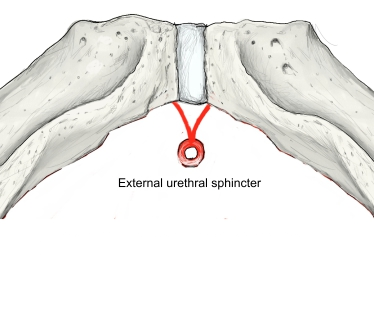
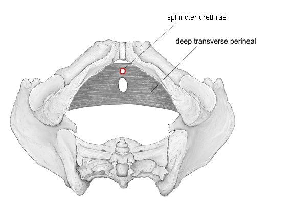
The illustration is schematic; it shows the general arrangement of the sphincter rather than its exact shape.
Illustrations
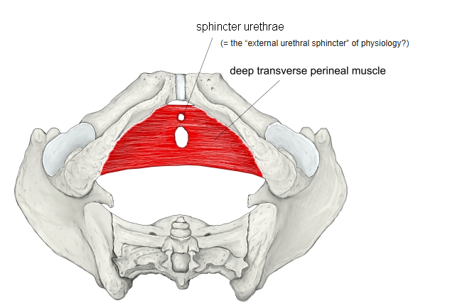
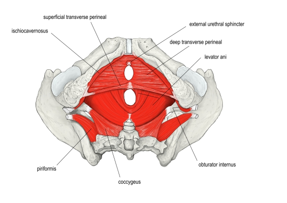
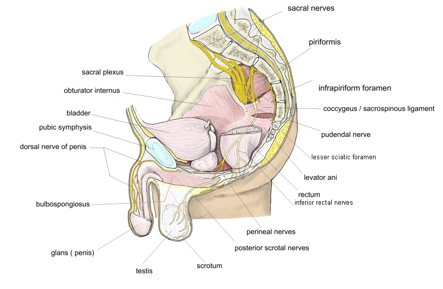
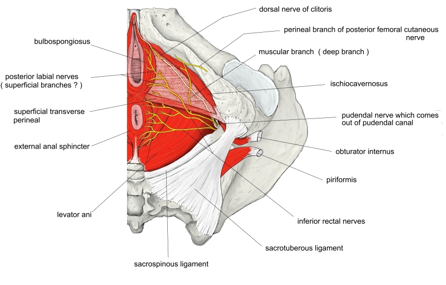
Origin / insertion
- Origin: inferior rami of the ischium and pubis; perineal body
- Insertion: fibres encircle the urethra and meet those of the other side, forming a ring
Action
- Closes the urethra voluntarily and controls urination
Innervation
- Perineal branch of the pudendal nerve (S2–S4); under voluntary control
Blood supply
- Internal pudendal artery, including its artery of the bulb of the penis (male) or of the vestibule (female)
Related terms
- Internal urethral sphincter
- Deep transverse perineal
- Bulbospongiosus
- Perineal muscles
- Urethra
References and sources
- Further reading: Urethral sphincters, Wikipedia.