Latin: musculus obliquus internus abdominis
Overview
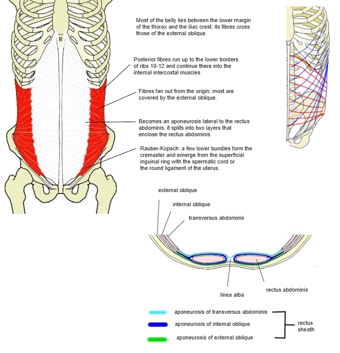
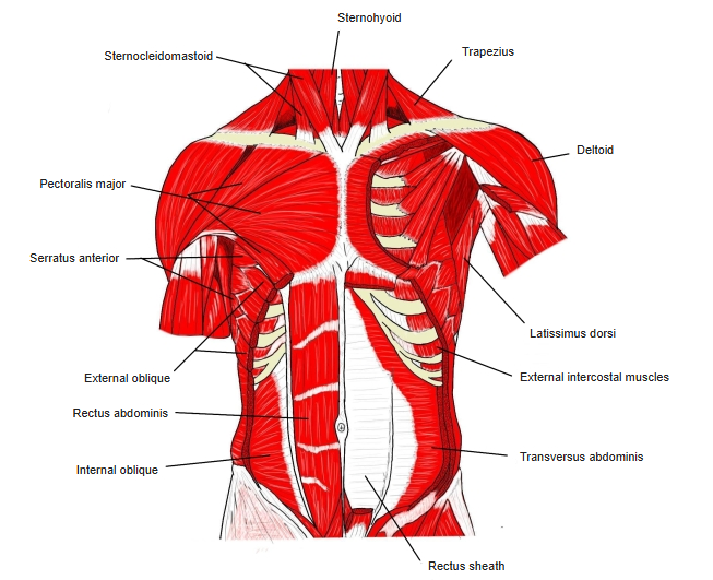
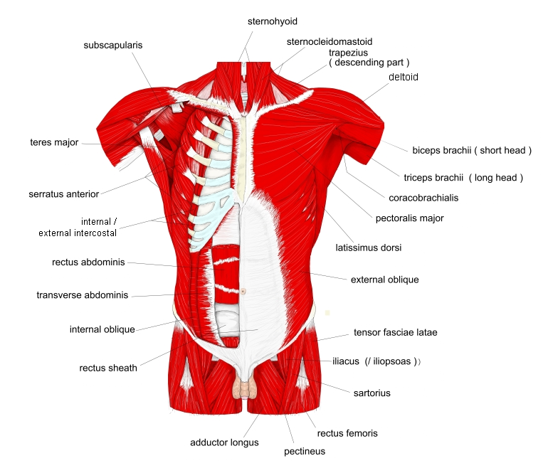
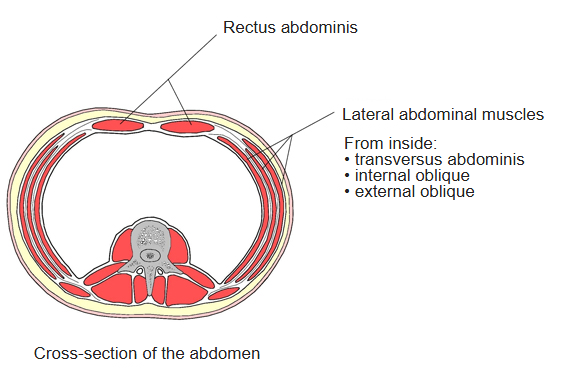
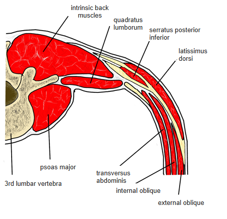
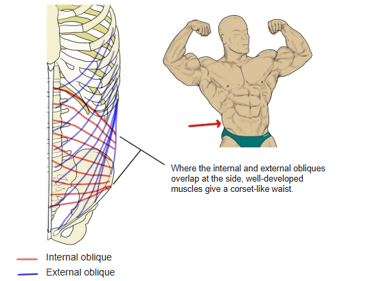
- The middle of the three flat muscles of the lateral abdominal wall; its fibres cross those of the external oblique.
- The three layers together are about 2 cm thick; the internal oblique is the thickest.
- Its aponeurosis interlaces at the linea alba with that of the external oblique of the other side, so the two obliques act as one unit.
- Upper fibres continue into the internal intercostal muscles; lower fibres are hard to separate from the transversus abdominis.
- A few lower bundles form the cremaster.
- Continuous in places with the external oblique, transversus abdominis and rectus abdominis.
Position in the classification
Trunk › Abdomen › Lateral abdominal muscles
- External oblique
- Internal oblique
- Transversus abdominis
Illustrations
Origin / insertion
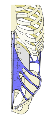
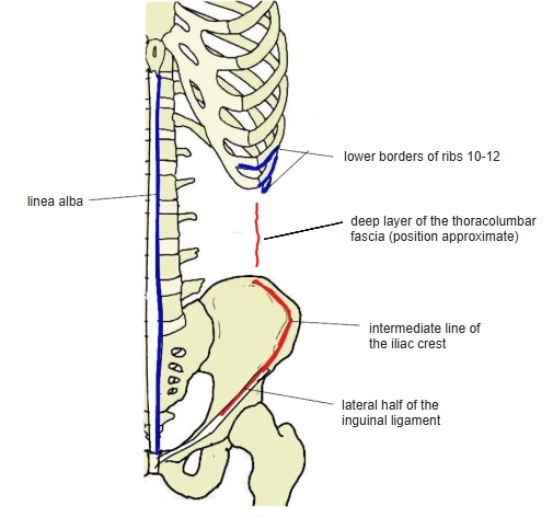
- Origin: lateral half of the inguinal ligament, intermediate line of the iliac crest, posterior layer of the thoracolumbar fascia
- Insertion: lower borders of ribs 10–12, rectus sheath, linea alba and (with the transversus abdominis, as the conjoint tendon) the pubic crest and medial part of the pectineal line
Action
- Pulls the thorax down; flexes the trunk, bends it to the same side and rotates it to the same side
- With the thorax fixed, raises the pelvis
- Compresses the abdomen; active in forced expiration
Innervation
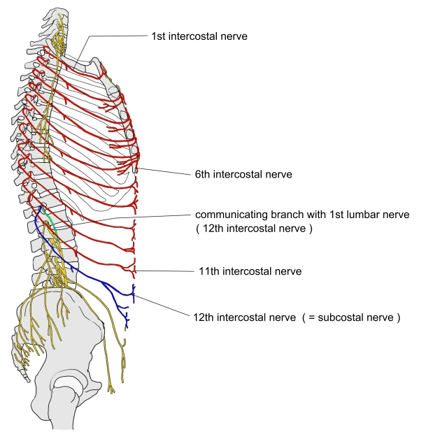
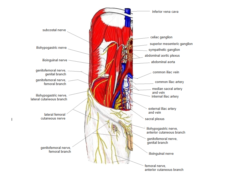
- Lower intercostal nerves (T7–T11) and the subcostal nerve (T12); also the iliohypogastric and ilioinguinal nerves (L1)
Blood supply
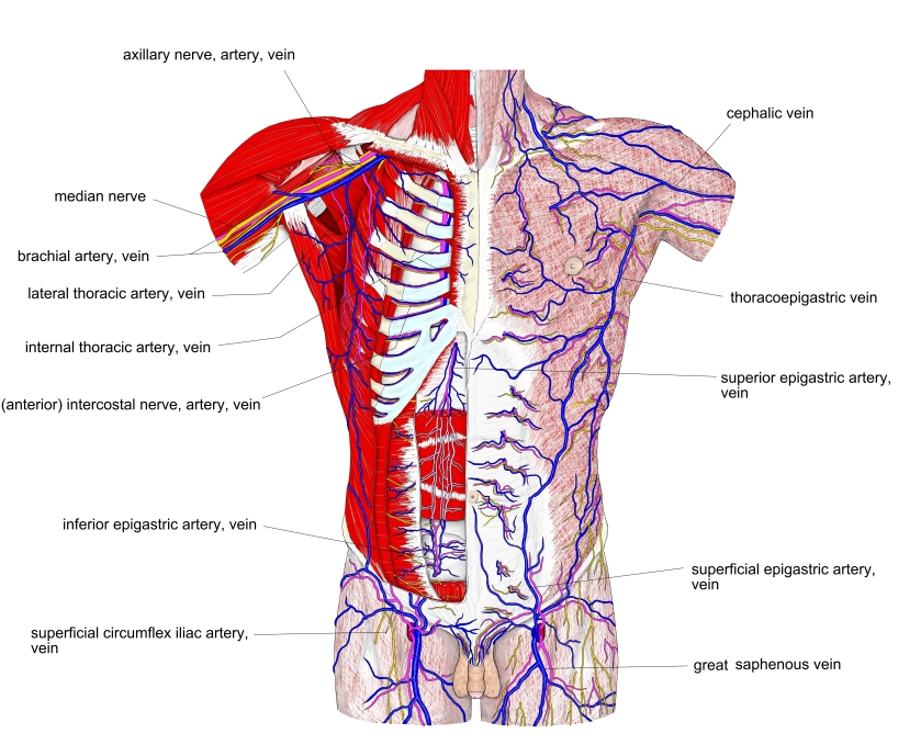
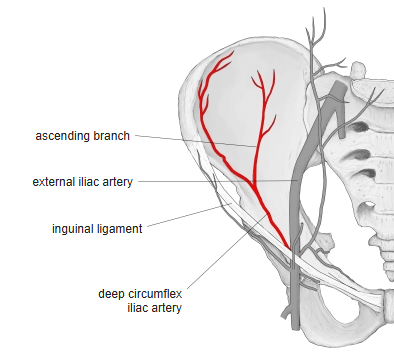
- Muscular branches of the superior and inferior epigastric arteries and the deep circumflex iliac artery
Anatomical variations
- The number of rib insertions may drop to two or rise to four; a tendinous intersection, sometimes with cartilage, often continues the lowest rib
Related terms
- External oblique
- Transversus abdominis
- Rectus abdominis
- Cremaster
- Linea alba
- Inguinal ligament
- Iliohypogastric nerve
References and sources
- Further reading: Abdominal internal oblique muscle, Wikipedia.