Latin: musculus transversus abdominis
Overview
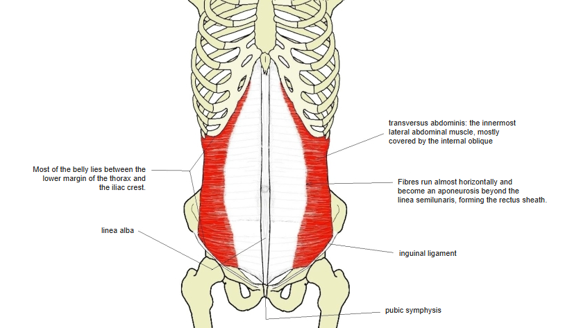
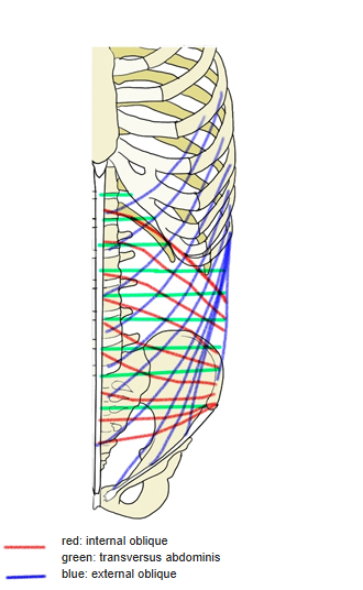
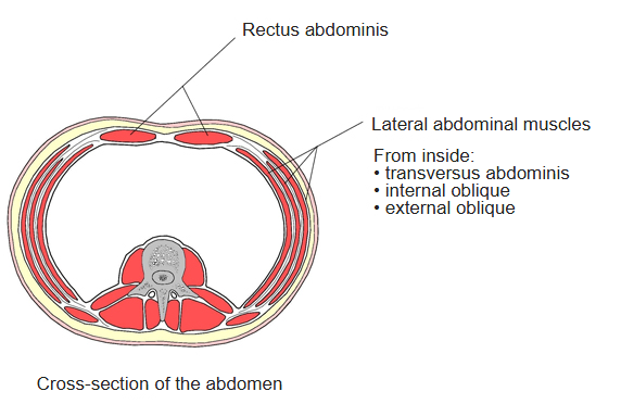
- The innermost of the flat lateral abdominal muscles, mostly covered by the internal oblique; a deep "core" muscle, like a corset.
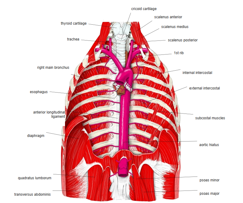
- Lies just below the transversus thoracis.
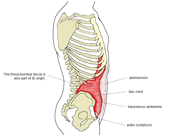
- Its fibres run almost horizontally and become an aponeurosis beyond the curved linea semilunaris, helping to form the rectus sheath.
- Most of the belly lies between the lower margin of the thorax and the iliac crest.
- Continuous in places with the internal oblique, rectus abdominis and diaphragm.
Position in the classification
Trunk › Abdomen › Lateral abdominal muscles
- External oblique
- Internal oblique
- Transversus abdominis
Illustrations
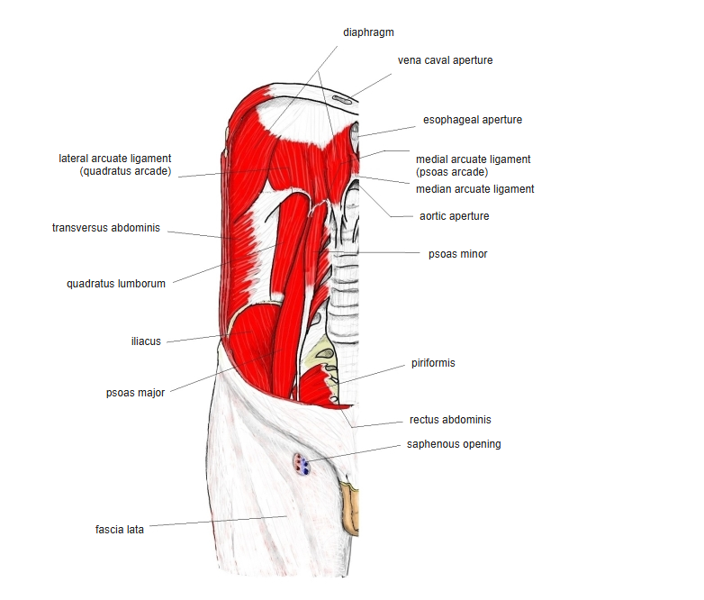
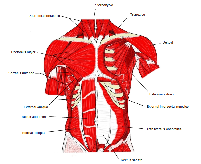
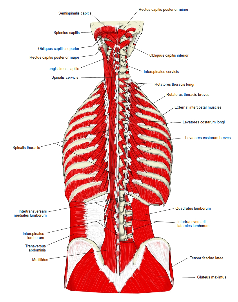
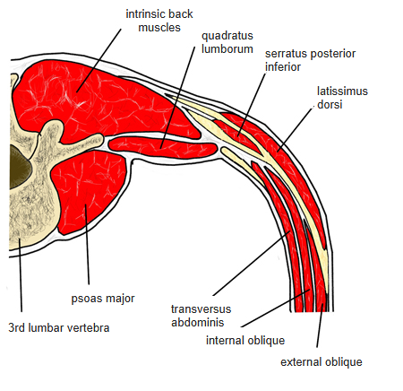
Origin / insertion
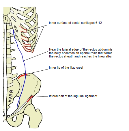
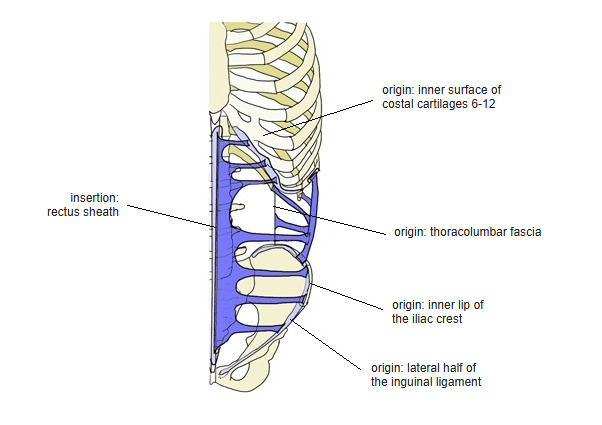
- Origin: inner surface of the 6th (or 7th) to 12th costal cartilages, thoracolumbar fascia, inner lip of the iliac crest, lateral half of the inguinal ligament
- Insertion: becomes an aponeurosis at the linea semilunaris and joins the rectus sheath, ending in the linea alba
Action
- Pulls down ribs 6–12 and raises abdominal pressure
- Compresses the abdomen and protects its organs; active in forced expiration
Innervation
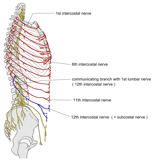
- Intercostal nerves (T7–T12), iliohypogastric and ilioinguinal nerves; some sources add the genitofemoral nerve
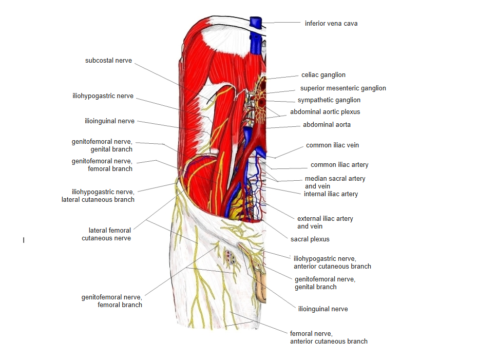
Blood supply
- Deep circumflex iliac artery, inferior epigastric artery
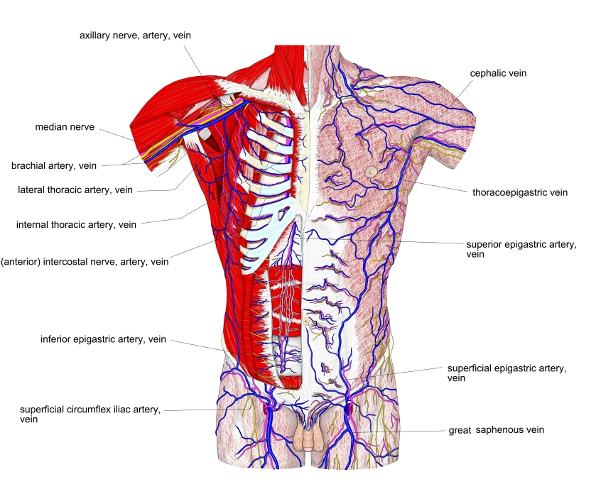
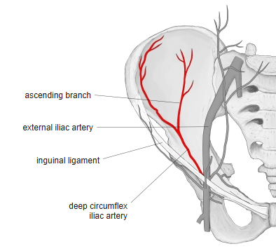
Anatomical variations
- May be completely absent; doubling known in one case; often arises from only the lower five ribs; connections with the internal oblique, internal intercostals, transversus thoracis and diaphragm (Rauber-Kopsch)
Related terms
- Internal oblique
- External oblique
- Rectus abdominis
- Linea alba
- Inguinal ligament
- Iliohypogastric nerve
References and sources
- Further reading: Transverse abdominal muscle, Wikipedia.