Latin: musculus opponens pollicis
Overview
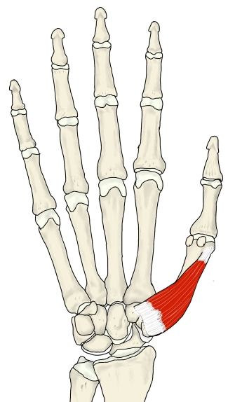
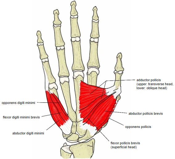
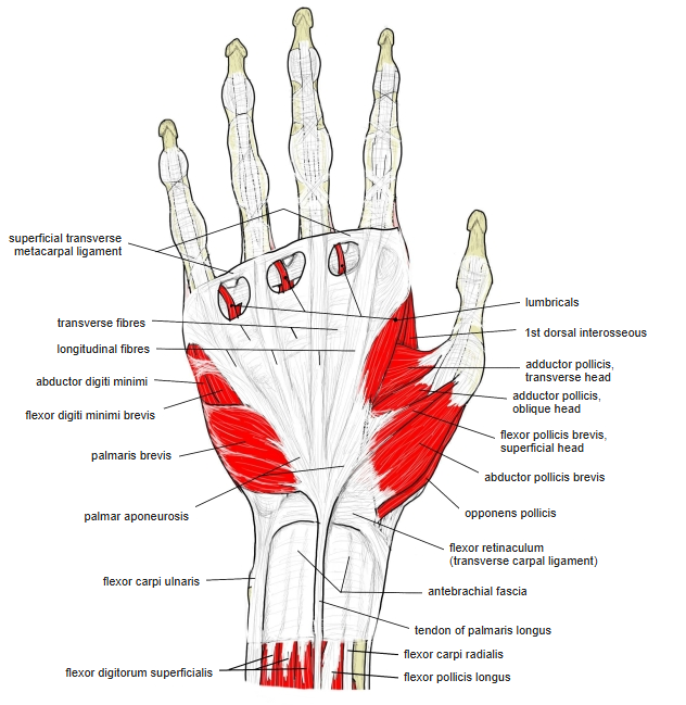
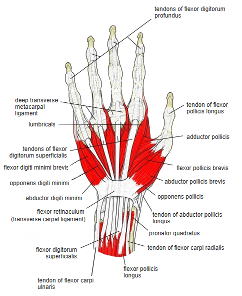
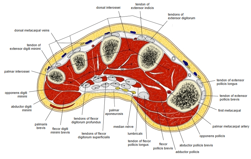
- A small triangular thenar muscle; most of its belly lies under the abductor pollicis brevis.
- Crosses under the abductor pollicis brevis at a sharp angle to reach the 1st metacarpal.
- Hard to feel on its own: the whole thenar eminence tightens when the thumb and little finger press together.
Position in the classification
Upper Limb › Hand › Thenar muscles
- Adductor pollicis
- Abductor pollicis brevis
- Flexor pollicis brevis
- Opponens pollicis
Illustrations
Origin / insertion
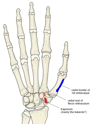
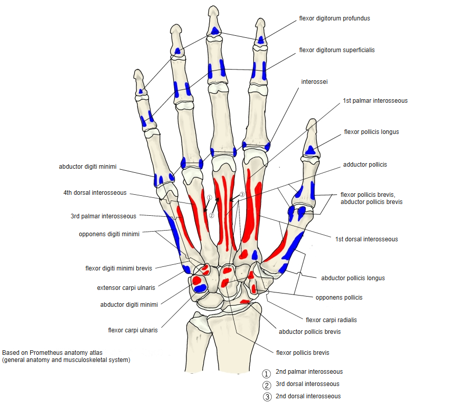
- Origin: tubercle of the trapezium and radial end of the flexor retinaculum
- Insertion: radial border of the 1st metacarpal (whole length)
Action
- Opposes the thumb (brings it towards the base of the little finger); some sources describe flexion of the thumb or of its carpometacarpal joint
- Helps adduct the thumb
Innervation
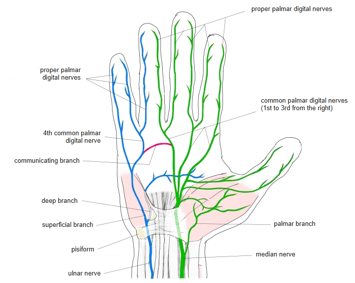
- Median nerve; sources give C8–T1, C6–T1, C6–C7 or C6–C8
- In about 20% supplied by the ulnar nerve (Wikipedia)
Blood supply
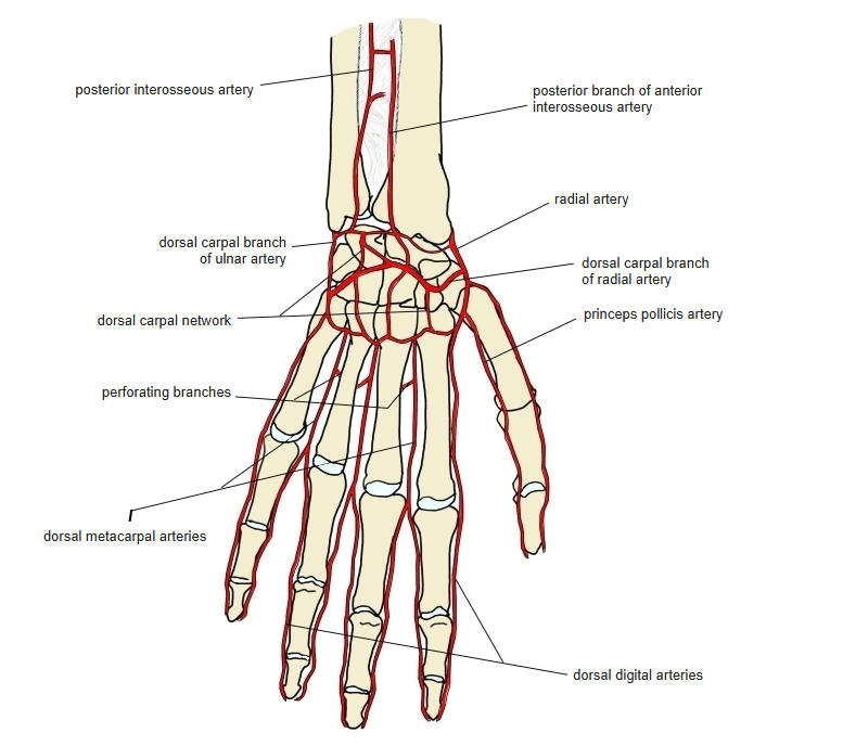
- Superficial palmar arch (branch of the ulnar artery; Wikipedia)
Related terms
- Abductor pollicis brevis
- Flexor pollicis brevis
- Adductor pollicis
- Trapezium
- Flexor retinaculum
- Median nerve
References and sources
- Further reading: Opponens pollicis muscle, Wikipedia.