Latin: musculus orbicularis oculi
Overview
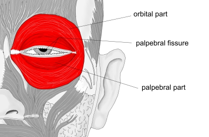
- A ring-shaped muscle around the palpebral fissure; the only muscle that closes the eye.
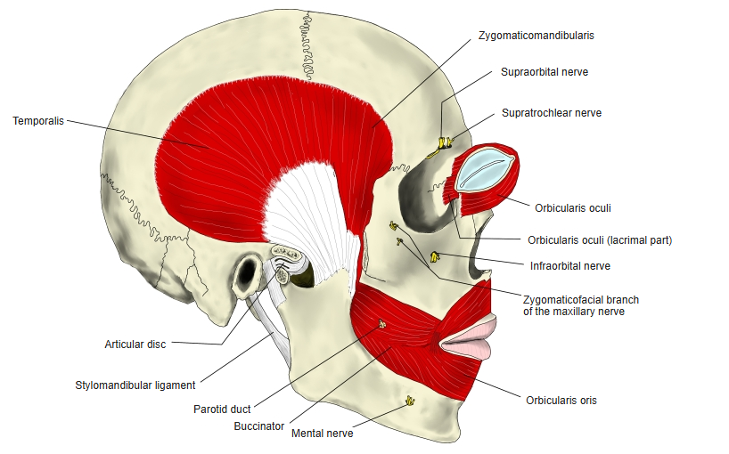
- Three parts: palpebral part (thin, pale, in the eyelids), orbital part (thicker, darker, around the orbit) and lacrimal part (small, deep, behind the lacrimal sac).
- Laterally it is not attached to bone.
- The eye is opened mainly by the levator palpebrae superioris, its antagonist.
- Variation: part of the orbital part may run into the orbit (transverse orbital muscle).
Position in the classification
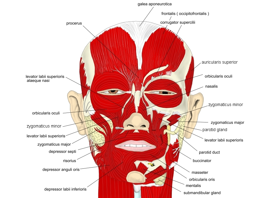
Head › Facial muscles › Muscles around the orbit and eyelids
- Orbicularis oculi
- Corrugator supercilii
- Depressor supercilii
Illustrations
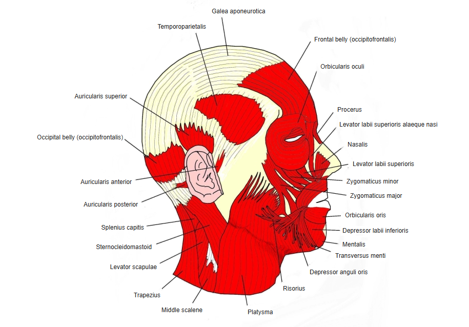
Origin / insertion
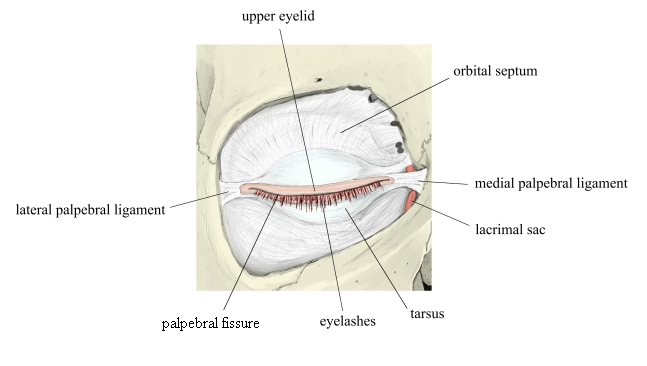
- Palpebral part: from the medial palpebral ligament to the lateral palpebral ligament (raphe)
- Orbital part: from the nasal part of the frontal bone, the anterior lacrimal crest of the maxilla and the medial palpebral ligament; runs round the orbital margin, partly to the lateral palpebral ligament or the skin of the eyebrow
- Lacrimal part: from the posterior lacrimal crest of the lacrimal bone; joins the palpebral part at the medial angle of the eye
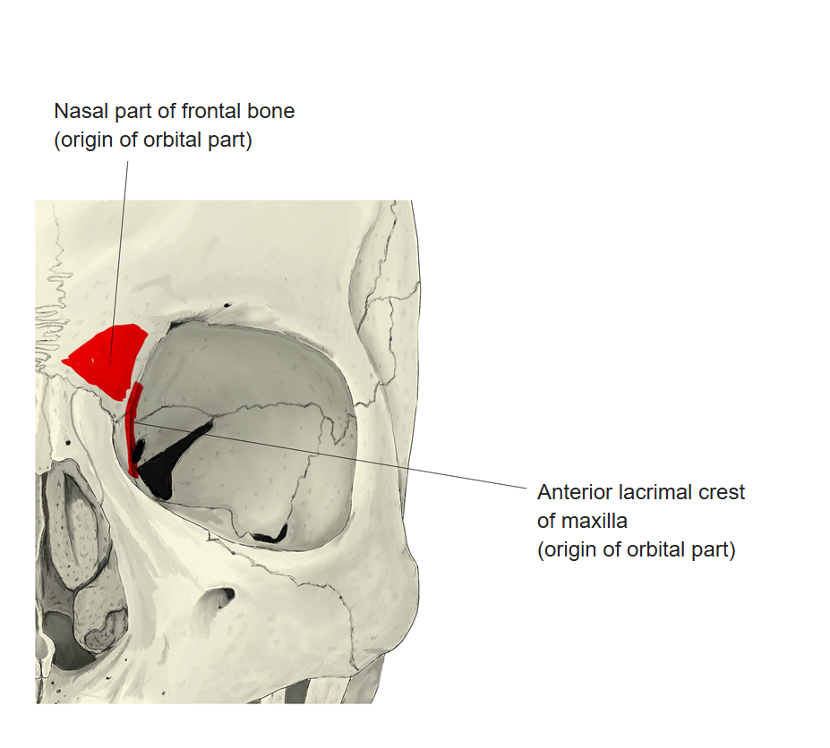
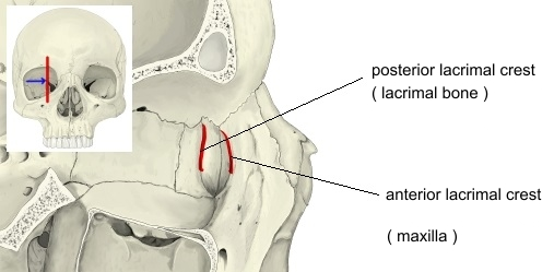
Action
- Closes the eyelids: the palpebral part gently and involuntarily (blinking, sleep), the orbital part firmly and voluntarily
- The lacrimal part helps to drain tears into the lacrimal sac
Innervation
- Facial nerve – temporal and zygomatic branches
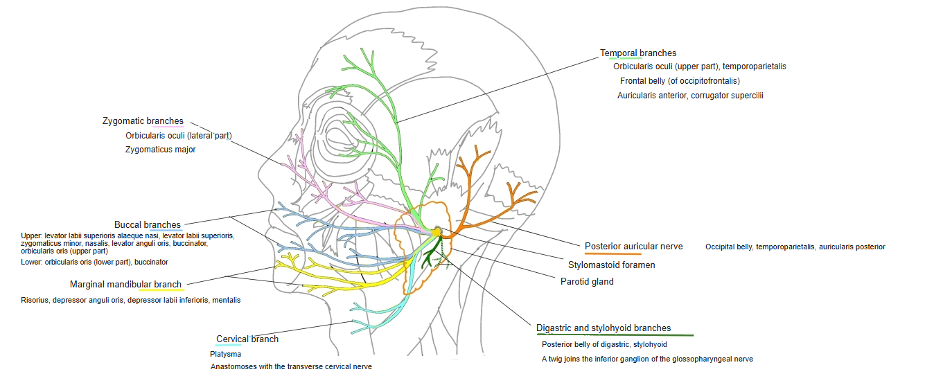
Blood supply
- Branches of the ophthalmic artery, the zygomatico-orbital artery and the angular artery
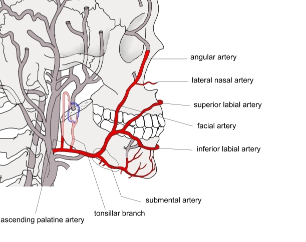
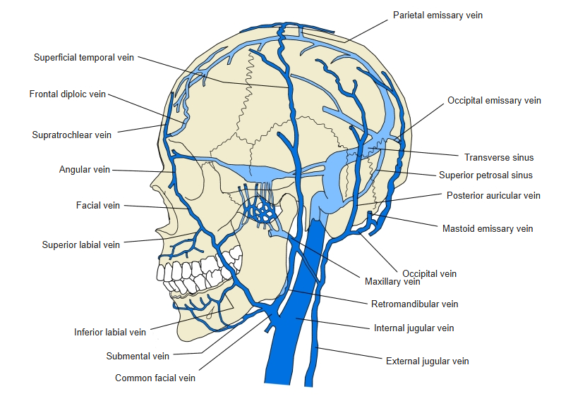
The arteries above are from Wikipedia.
Related terms
- Levator palpebrae superioris
- Corrugator supercilii
- Frontal belly (occipitofrontalis)
- Transverse orbital muscle
- Facial nerve
References and sources
- Further reading: Orbicularis oculi muscle, Wikipedia.