Latin: musculus plantaris
Overview
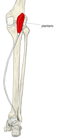
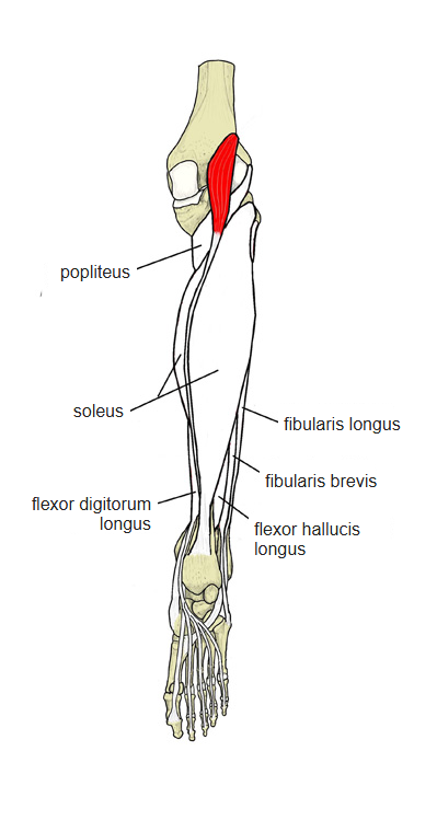
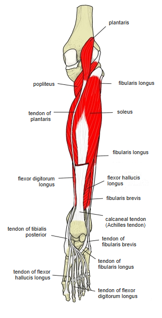
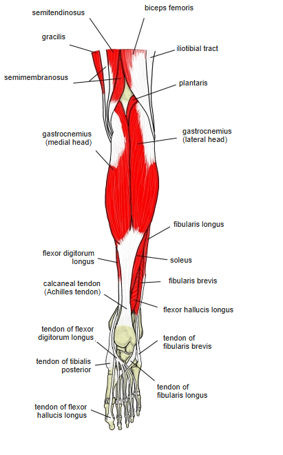
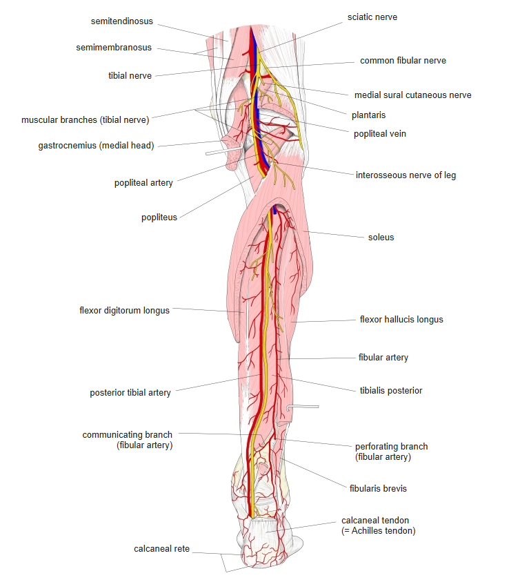
- A small muscle with a short, thin belly and a very long, thin tendon, lying between the gastrocnemius and the soleus in a sleeve of connective tissue.
- Its tendon is said to be the longest in the body (30–45 cm; Wikipedia); the belly is only about 5–10 cm long.
- A phylogenetic remnant of a muscle that continues into the plantar aponeurosis in lower primates.
- Sometimes absent (8–12%) or fused with the lateral head of the gastrocnemius; sometimes continues into the crural fascia, rarely into the plantar aponeurosis.
- Its connective tissue is linked with the sheath of the posterior tibial vessels; above the tendinous arch of the soleus it runs over these vessels.
Position in the classification
Lower Limb › Leg › Flexor group › Others
Illustrations
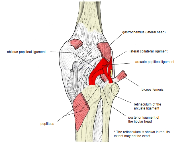
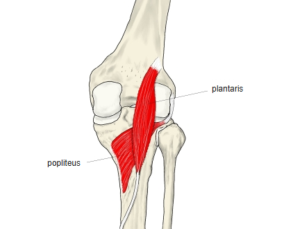
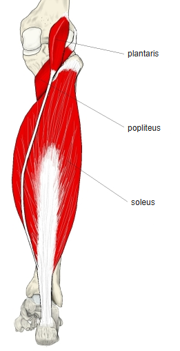
Origin / insertion
- Origin: lateral epicondyle of the femur (lateral supracondylar line) and the knee joint capsule, especially the arcuate popliteal ligament
- Insertion: calcaneal tuberosity by way of the calcaneal (Achilles) tendon
- Also into the crural fascia and the plantar aponeurosis (some sources)
- Sometimes a separate tendon ends on the calcaneus (Wikipedia)
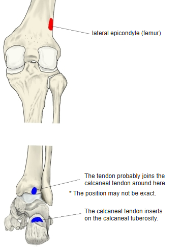
Action
- Assists the triceps surae: weak plantar flexion at the ankle and weak flexion of the knee
- A vestigial muscle; its own action is weak
- During knee flexion it may lift the neurovascular bundle into an arch so that the vessels do not kink
Innervation
- Tibial nerve (L4–S1)
Blood supply
- Sural arteries, fibular artery
Related terms
- Gastrocnemius
- Soleus
- Popliteus
- Calcaneal tendon (Achilles tendon)
- Femur
- Calcaneus
- Tibial nerve
References and sources
- Further reading: Plantaris muscle, Wikipedia.