Latin: musculus tibialis posterior
Overview
- A muscle of the flexor group of the leg (one of the "other" deep flexors, not the triceps surae).
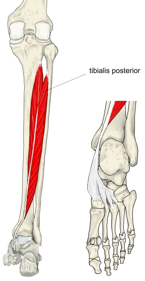
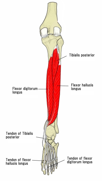
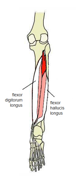
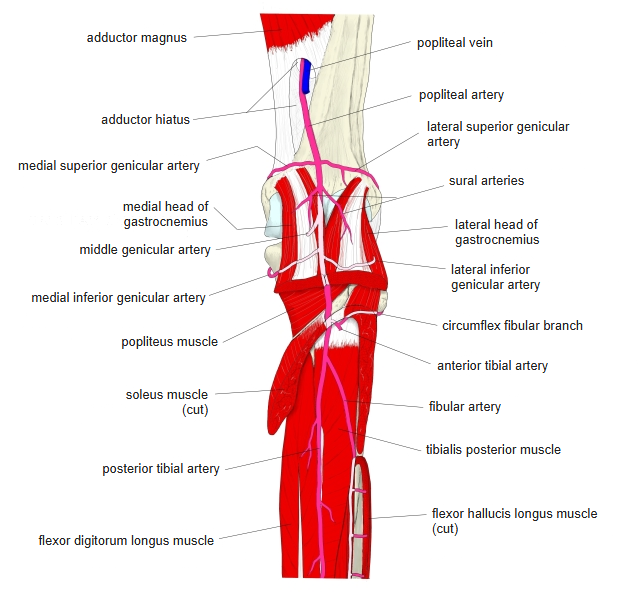
- Lies in the middle of the deep layer at the back of the leg, deep to the soleus, on the interosseous membrane.
- Pennate (the lower part is unipennate).
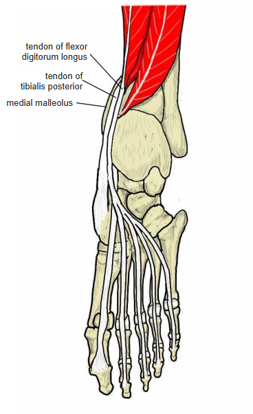
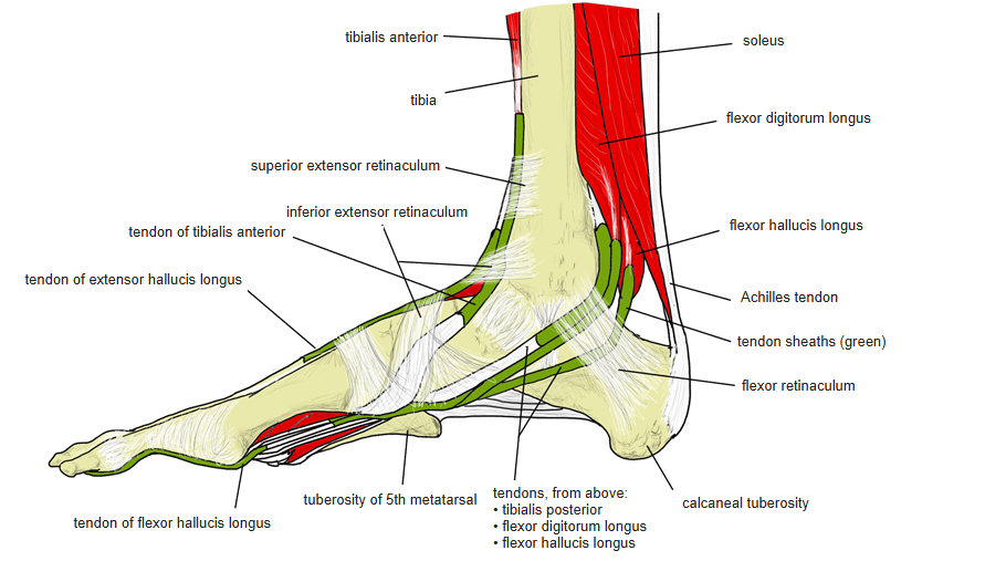
- Above the medial malleolus its tendon crosses under the tendon of the flexor digitorum longus from lateral to medial (the crural chiasma).
- One of the muscles that support the arch of the foot, especially the medial longitudinal arch.
- Muscles connected with it: flexor digitorum longus, flexor hallucis longus, soleus, tibialis anterior, extensor hallucis longus and popliteus.
Position in the classification
Lower Limb › Leg › Flexor group › Others
- Flexor digitorum longus
- Flexor hallucis longus
- Tibialis posterior
- Popliteus
- Plantaris
Illustrations
Origin / insertion
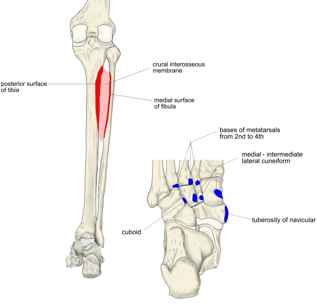
- Origin: posterior surface of the tibia, medial surface of the fibula and the interosseous membrane of the leg
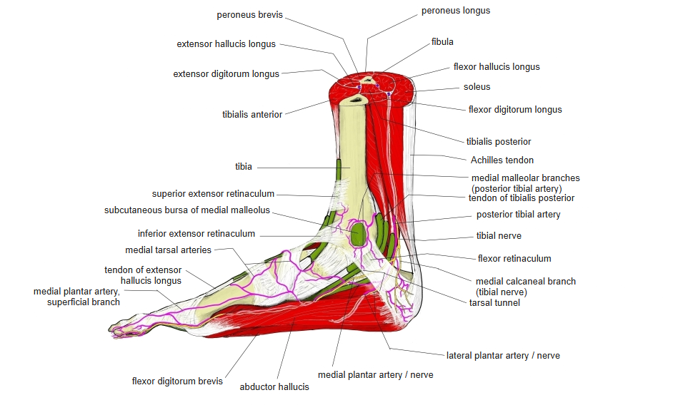
- The tendon descends behind the medial malleolus in a tendon sheath, covered below the malleolus by the flexor retinaculum.
- Insertion:
- Main part: tuberosity of the navicular
- Also the medial, intermediate and lateral cuneiforms, the cuboid and the plantar surface of the bases of the 2nd–4th metatarsals
- Some texts (e.g. the Prometheus atlas) do not include the cuboid.
Action
- Plantarflexes the foot and at the same time inverts it
- Supports the medial longitudinal arch and helps to draw the bones of the sole together
- Failure of the muscle or its tendon can lead to flat foot in adults.
Innervation
- Tibial nerve (L5, S1, S2)
- Its branch leaves the tibial nerve higher than the branches to the other deep flexors, at the level of the tendinous arch of the soleus.
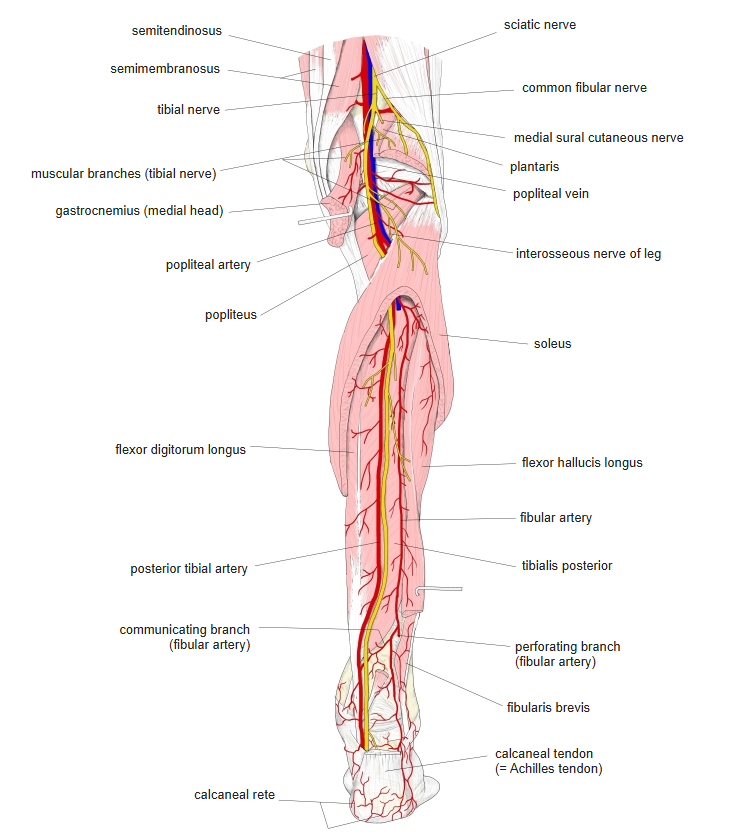
Blood supply
- Posterior tibial artery
- Fibular artery
Related terms
- Muscles of the leg
- Flexor digitorum longus
- Flexor hallucis longus
- Soleus
- Medial malleolus
- Tendon of tibialis posterior
- Tibial nerve
References and sources
- Further reading: Tibialis posterior muscle, Wikipedia.