Latin: musculus pronator teres
Overview
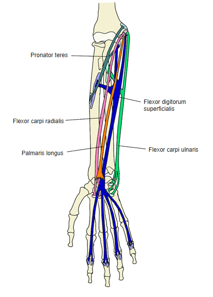
- A muscle of the proximal forearm in the first (superficial) layer of the forearm flexors.
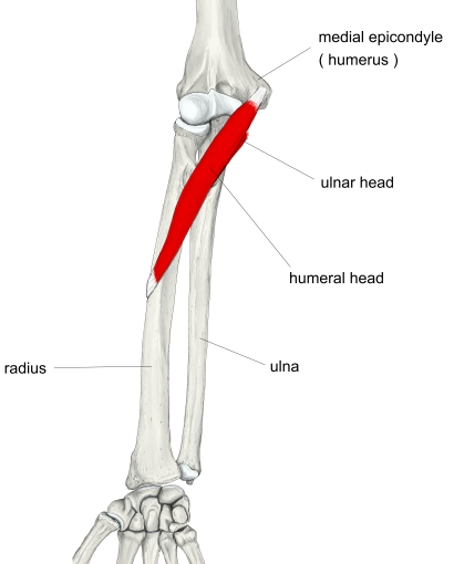
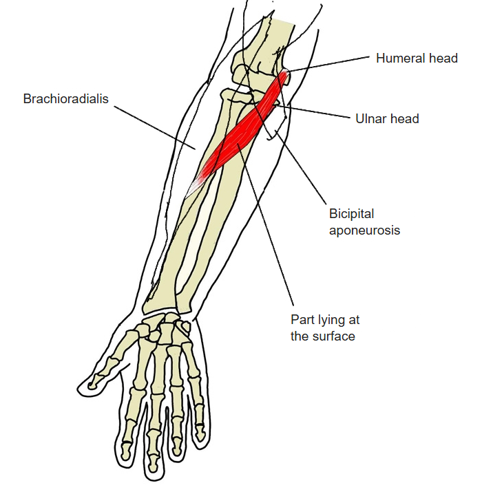
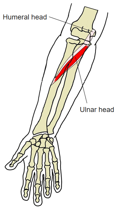
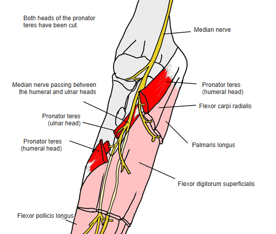
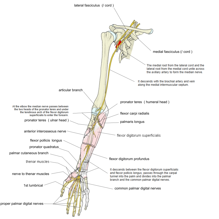
- Has two heads, humeral and ulnar, which join in a common belly; the median nerve passes between them.
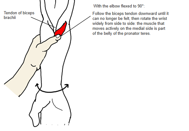
- Covered near its origin by the bicipital aponeurosis and near its insertion by the brachioradialis, so only the middle third lies at the surface and can be palpated.
- Connected with the flexor digitorum superficialis, flexor carpi radialis and biceps brachii; its lateral border forms the medial boundary of the cubital fossa.
Position in the classification
Upper Limb › Forearm › Flexor group › First layer
- Pronator teres
- Flexor carpi radialis
- Palmaris longus
- Flexor carpi ulnaris
Illustrations
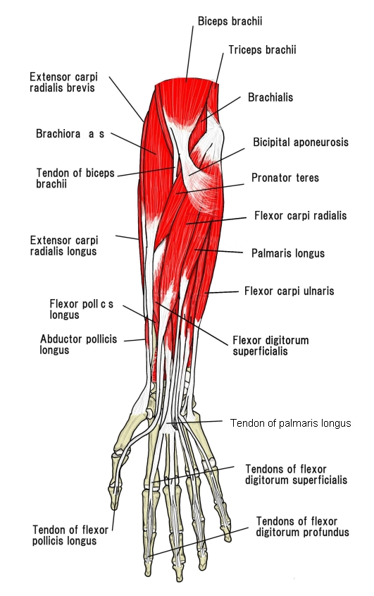
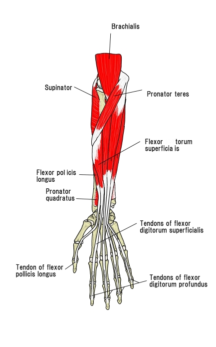
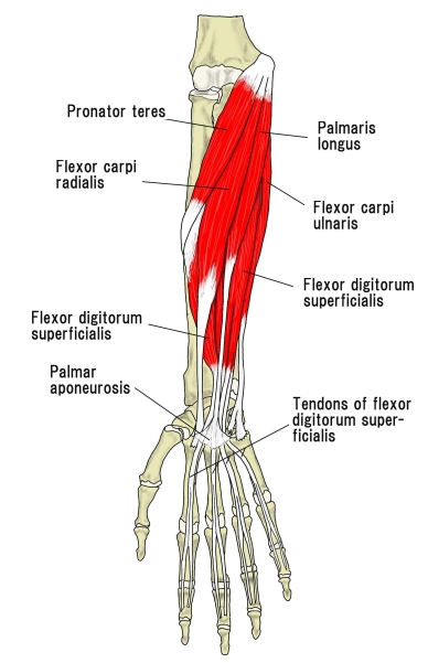
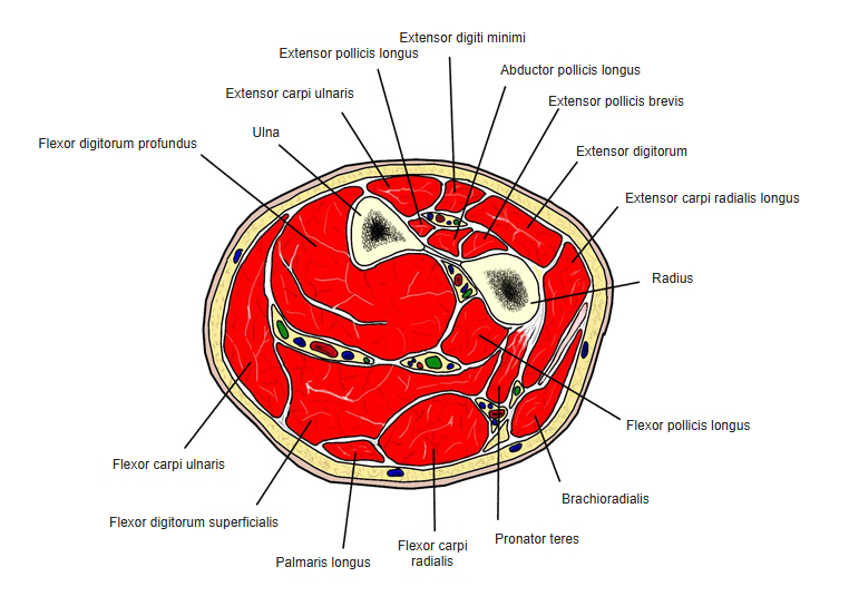
Origin
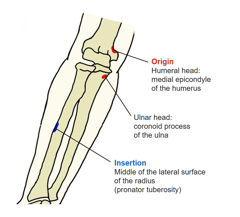
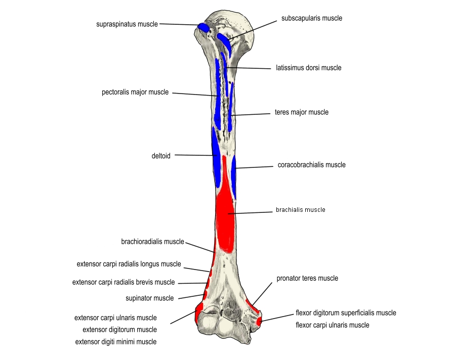
- Humeral head: medial epicondyle of the humerus (the medial supracondylar ridge just above it and the common flexor tendon); some sources also list the medial intermuscular septum
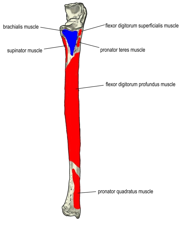
- Ulnar head: medial side of the coronoid process of the ulna
Insertion
- Middle of the lateral surface of the radius (pronator tuberosity), just distal to the insertion of the supinator
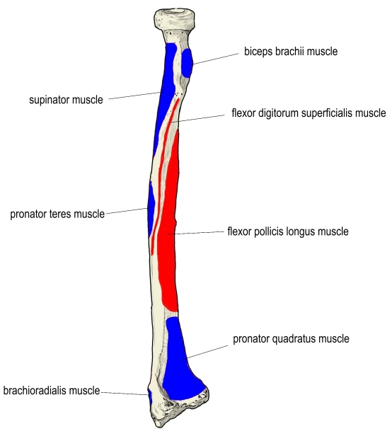
Action
- Pronation of the forearm, assisted by the pronator quadratus
- Weak flexion of the elbow
Innervation
- Median nerve (C6, C7), which enters the forearm between the two heads
Blood supply
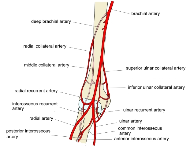
- Ulnar artery
- Superior and inferior ulnar collateral arteries (not certain)
Anatomical variations
- The ulnar head is often absent or rudimentary
- The two heads may be completely separate
- A third head may arise from the medial intermuscular septum and the supracondylar process
- A sesamoid bone is occasionally found at the origin of the humeral head
- May be joined to the brachialis, palmaris longus or flexor digitorum superficialis
Related terms
- Medial epicondyle
- Coronoid process
- Radius
- Pronator quadratus
- Supinator
- Flexor carpi radialis
- Palmaris longus
- Flexor carpi ulnaris
- Flexor digitorum superficialis
- Bicipital aponeurosis
- Brachioradialis
- Cubital fossa
- Median nerve
- Ulnar artery
References and sources
- Further reading: Pronator teres muscle, Wikipedia.