Latin: musculus psoas major
Overview
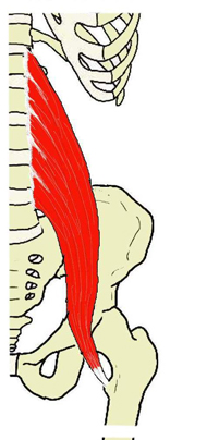
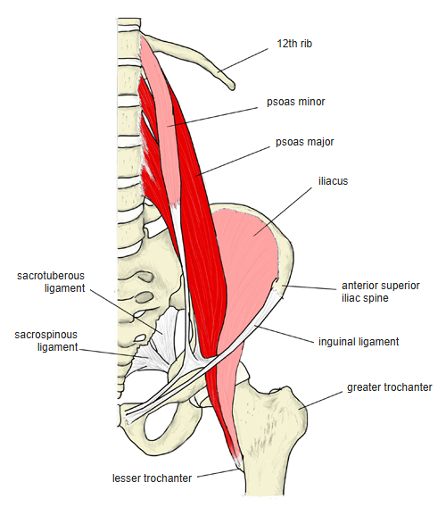
- A long, fusiform muscle beside the lumbar spine; it joins the iliacus to form the iliopsoas.
- Two heads, superficial and deep; several branches of the lumbar plexus run between them.
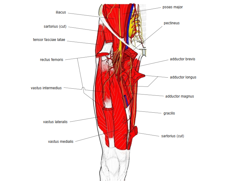
- Passes behind the medial arcuate ligament of the diaphragm; below, with the iliacus, it runs under the inguinal ligament through the muscular lacuna to the thigh.
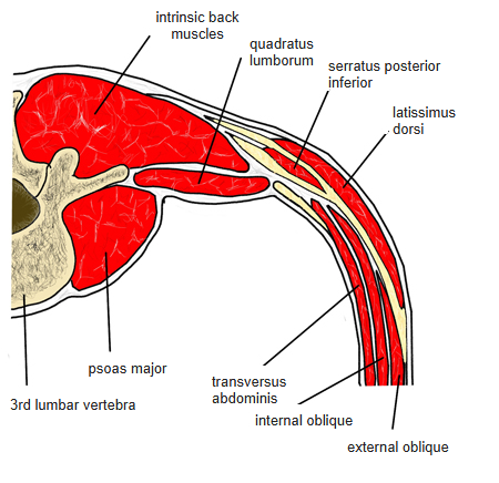
- Seen from the side it lies at about the middle of the trunk's front-to-back depth; its belly is thickest at the level of L4.
- Continuous in places with the iliacus, psoas minor, quadratus lumborum, diaphragm, longissimus and iliocostalis.
Position in the classification
Lower Limb › Hip › Inner hip muscles (iliopsoas)
- Iliacus
- Psoas major
- Psoas minor
Illustrations
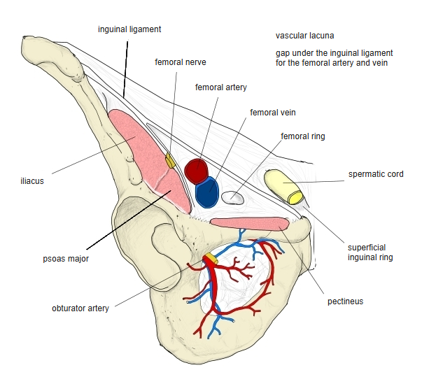
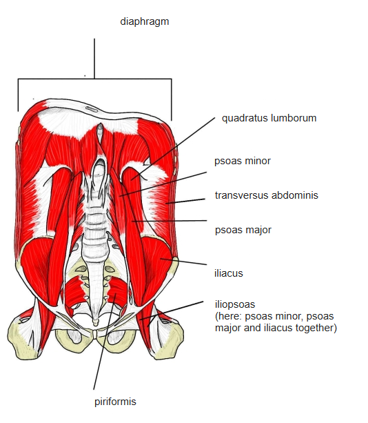
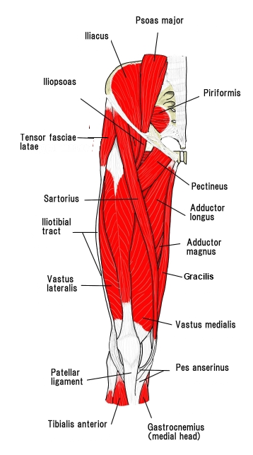
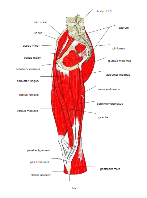
Origin / insertion
- Origin:
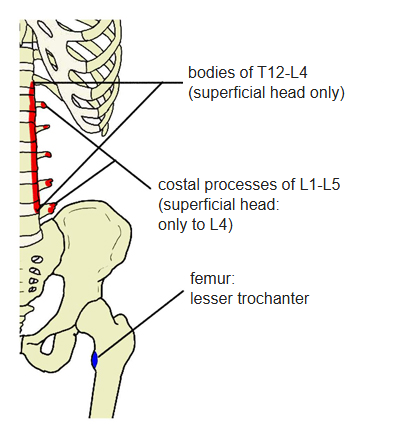
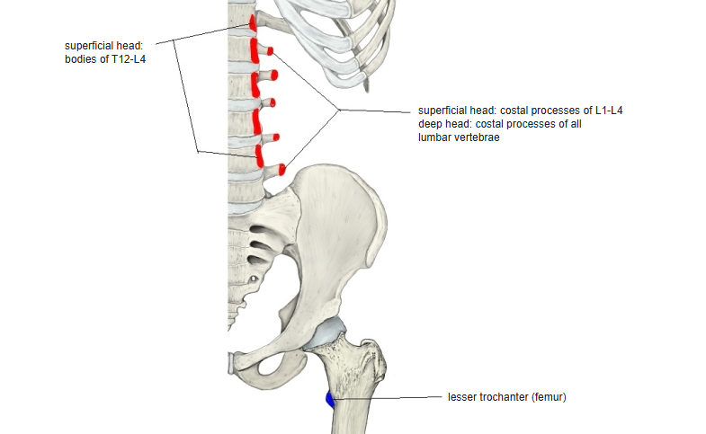
- Superficial head: bodies of T12–L4 (some sources add the intervertebral discs) and the costal processes of L1–L4
- Deep head: costal processes of all lumbar vertebrae
- Insertion: lesser trochanter of the femur, by a common tendon with the iliacus after passing through the muscular lacuna
Action
- Flexes the hip
- Laterally rotates the hip
- Pulls the lumbar spine forwards and downwards
Innervation
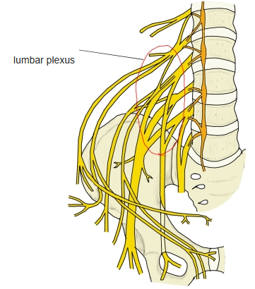
- Muscular branches of the lumbar plexus (L1–L3; sometimes T12 and L4)
Blood supply
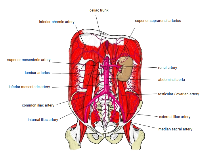
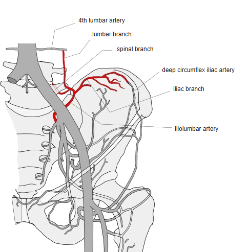
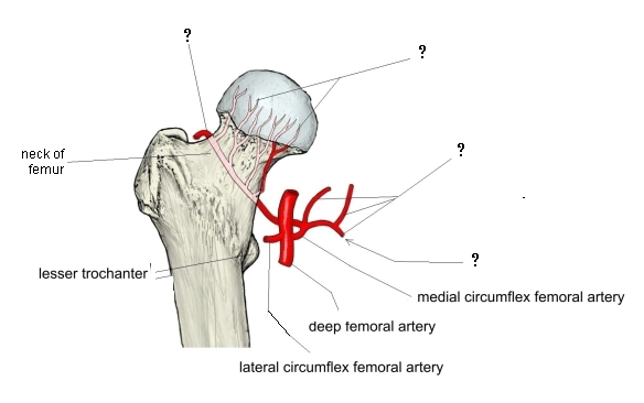
- Subcostal, lumbar and iliolumbar arteries, medial circumflex femoral artery
Related terms
- Iliacus
- Psoas minor
- Iliopsoas
- Quadratus lumborum
- Lesser trochanter
- Inguinal ligament
- Lumbar plexus
- Iliolumbar artery
References and sources
- Further reading: Psoas major muscle, Wikipedia.