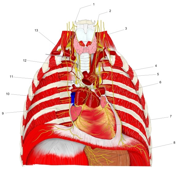
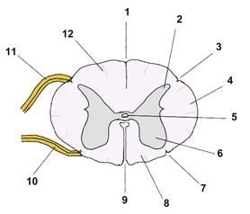
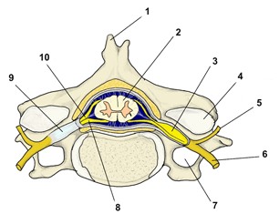
Numbered illustrations that show several nerves together, each with a table of names. Choose an illustration, then follow a name in the table to the page for that nerve. See also Nerve illustrations (one illustration for each nerve), List of nerves (A–Z) and Nerves by region.
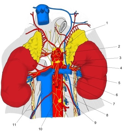
Brain and spinal cordCranial nervesSpinal nervesAutonomic nervous system
Brain and spinal cord
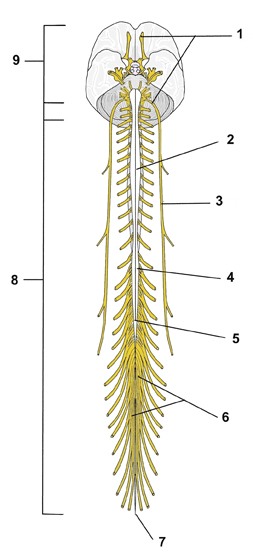
Cranial nerves
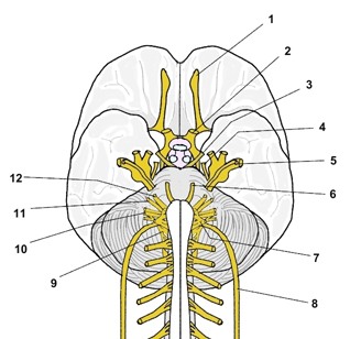 Cranial nerves I to XII on the base of the brain (schematic)
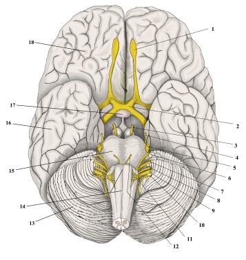 Cranial nerves on the base of the brain (inferior view, overview)
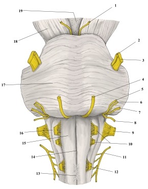 Cranial nerves at the brainstem (anterior view, enlarged)
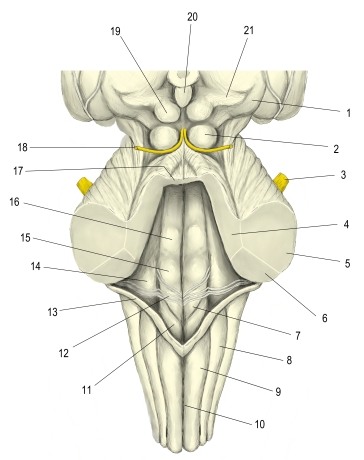 Brainstem with cerebellum removed (posterior view)
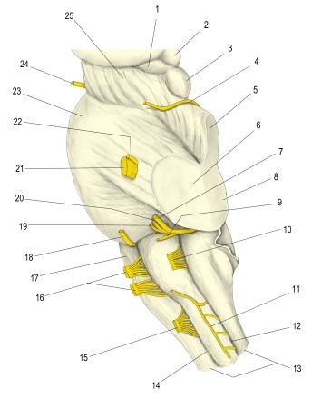 Cranial nerves and brainstem, cerebellum removed (left lateral view)
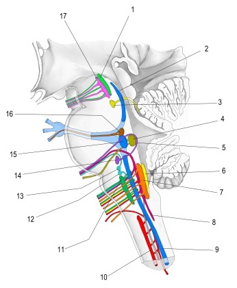 Cranial nerve nuclei in the brainstem (lateral view)
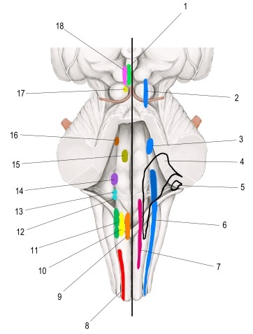 Cranial nerve nuclei (projection on brainstem)
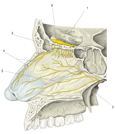 Nerves of the nasal septum (left lateral view)
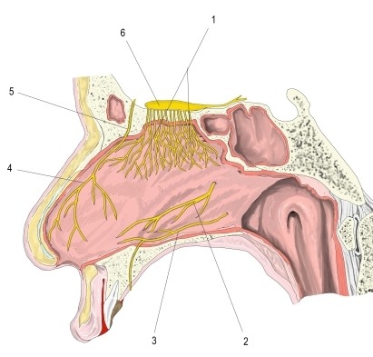 Nerves of the nasal septum, mucosa (left lateral view)
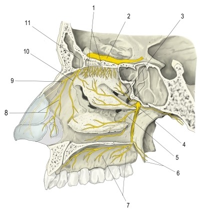 Nerves of the lateral wall of the right nasal cavity
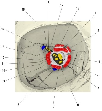 Superior orbital fissure, left (anterior view)
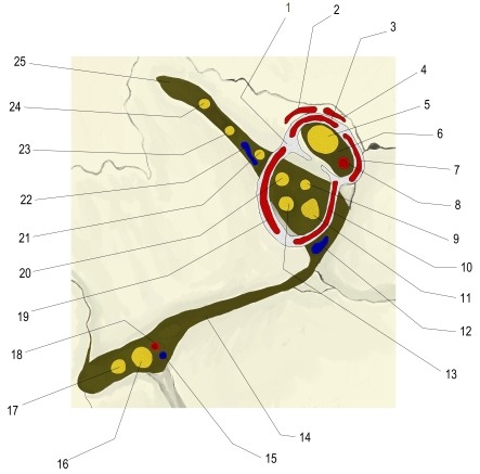 Orbital fissures and common tendinous ring (anterior view)
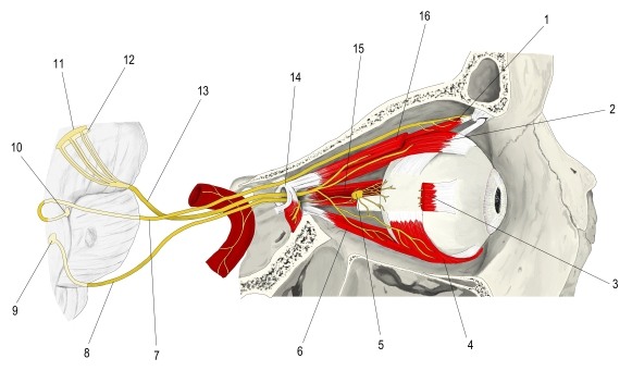 Oculomotor nerve and nuclei, right orbit (lateral view)
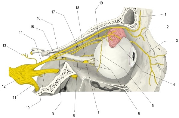 Ophthalmic nerve in the orbit (lateral view, sagittal section)
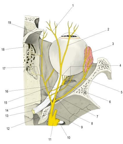 Ophthalmic nerve in the right orbit (superior view, horizontal section)
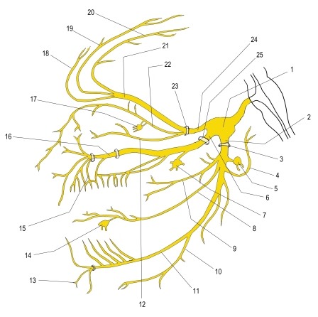 Trigeminal nerve and its three divisions (schematic)
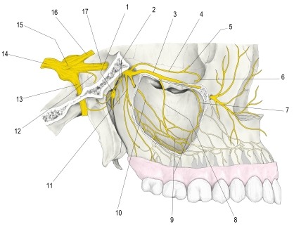 Maxillary nerve (lateral view)
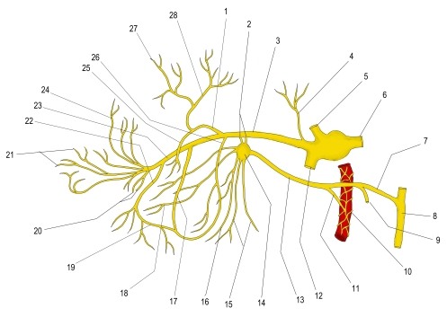 Maxillary nerve, branches (schematic)
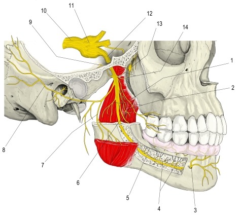 Mandibular nerve (lateral view)
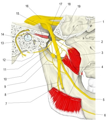 Mandibular nerve and otic ganglion (medial view)
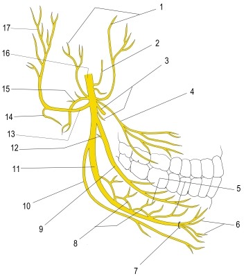 Mandibular nerve, branches (schematic)
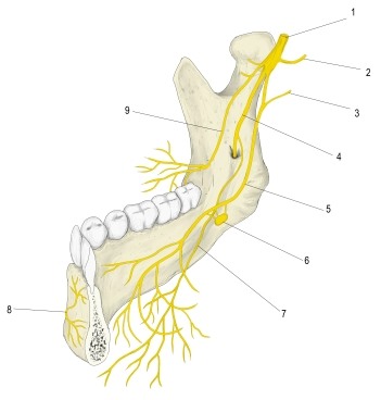 Mandibular nerve and its branches
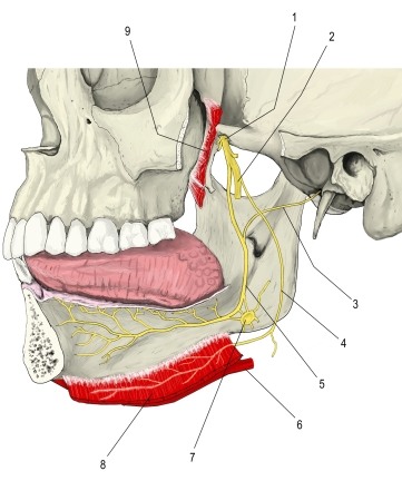 Lingual nerve and submandibular ganglion (lateral view, mandible removed)
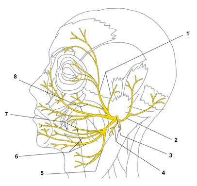 Facial nerve, branches in the face (lateral view)
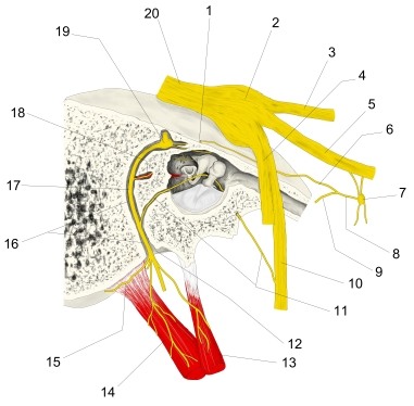 Facial nerve in the petrous part of the right temporal bone (lateral view)
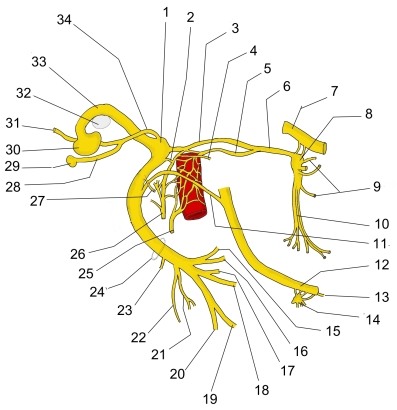 Facial nerve (schematic)
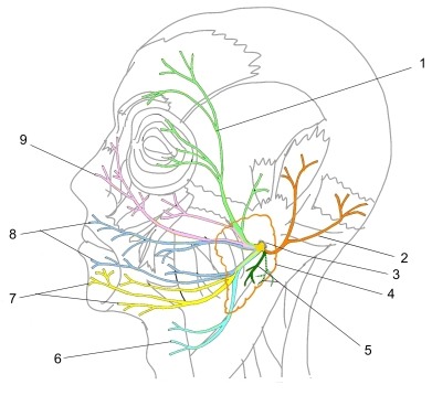 Facial nerve, branches after the stylomastoid foramen
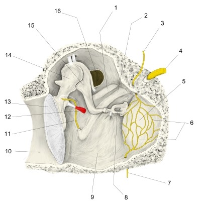 Walls of tympanic cavity (anterior wall removed)
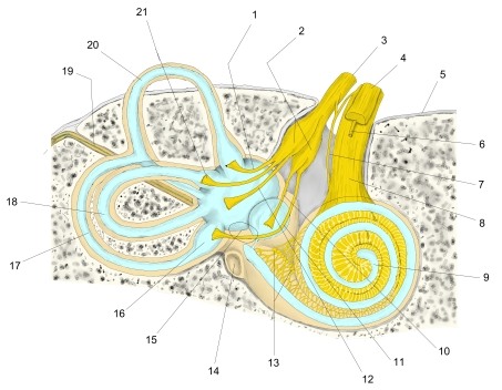 Vestibulocochlear nerve and inner ear in the temporal bone, right ear (anterior view)
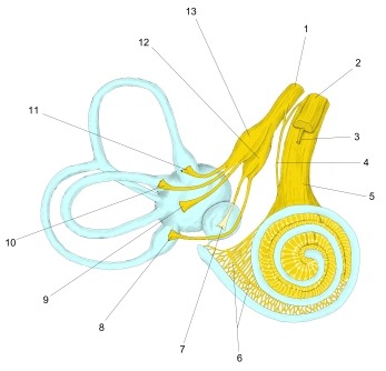 Vestibulocochlear nerve and membranous labyrinth, right ear (anterior view, simplified)
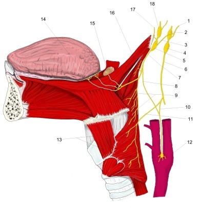 Glossopharyngeal nerve (schematic)
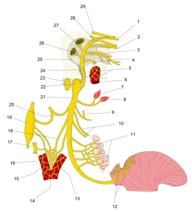 Right glossopharyngeal nerve (schematic)
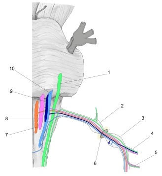 Vagus nerve, nuclei of origin (anterior view, schematic)
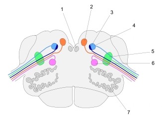 Vagus nerve, nuclei of origin (section at level of olive)
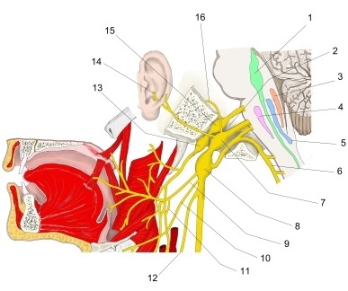 Vagus nerve, head and neck, with brainstem nuclei
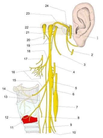 Vagus nerve, head and neck (schematic)
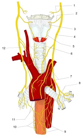 Vagus nerve, neck and thorax (anterior view, schematic)
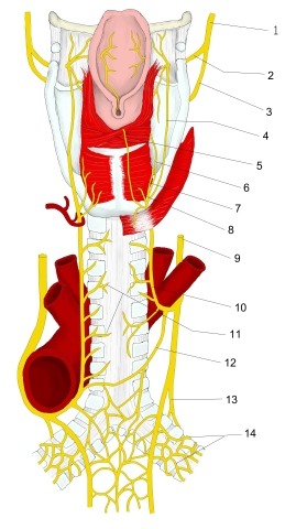 Vagus nerve, head and neck (schematic, posterior view)
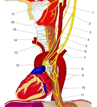 Vagus nerve, neck and thorax, branches (schematic)
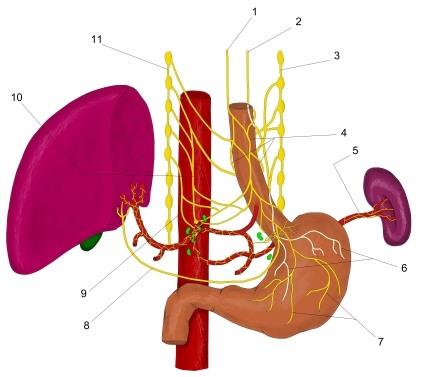 Vagus nerve, thorax and abdomen (schematic)
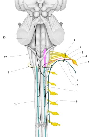 Accessory nerve, roots and nuclei of origin (schematic, posterior view)
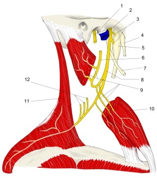 Accessory nerve, external branch (schematic, right lateral view)
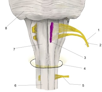 Hypoglossal nerve and its nucleus (schematic)
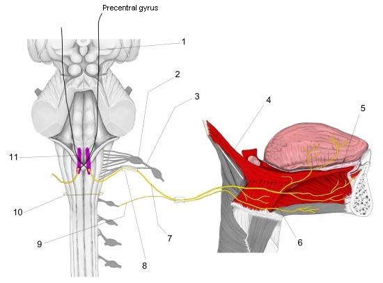 Hypoglossal nerve, nucleus and course (schematic I)
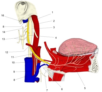 Hypoglossal nerve (schematic II)
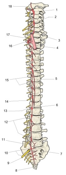
Spinal nerves
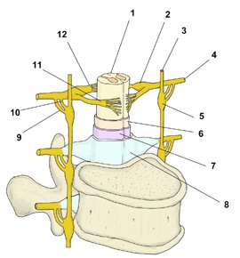
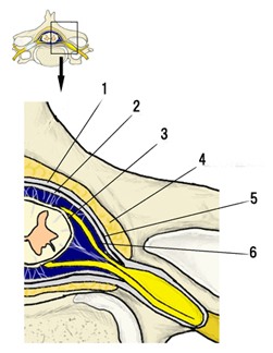
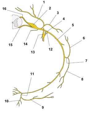 Typical spinal nerve: anterior and posterior rami (schematic)
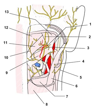 Cervical nerves, suboccipital region (posterior view)
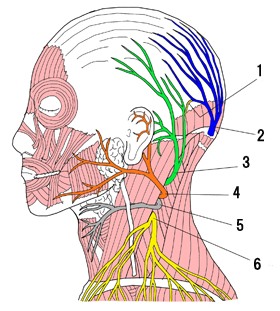 Cervical plexus, cutaneous branches (lateral view)
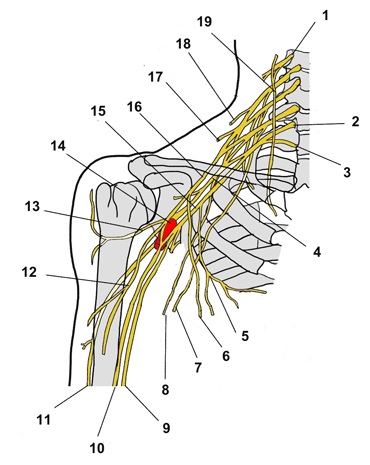 Cervical and brachial plexuses, right side (anterior view)
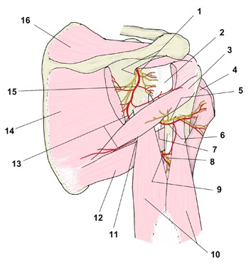 Scapular region, nerves and arteries (posterior view)
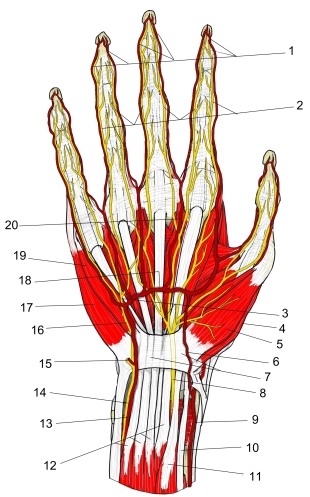 Main nerves and arteries of the palm (right hand)
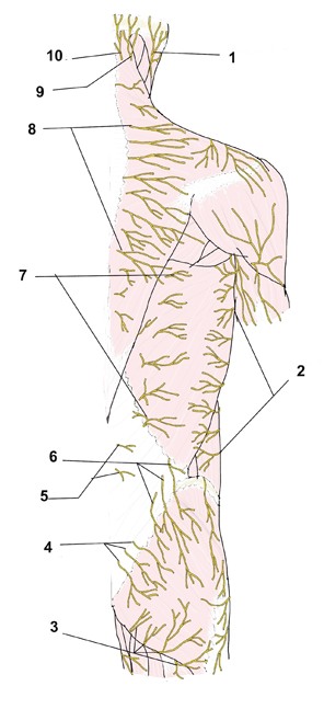 Nerves of the back, cutaneous branches (posterior view)
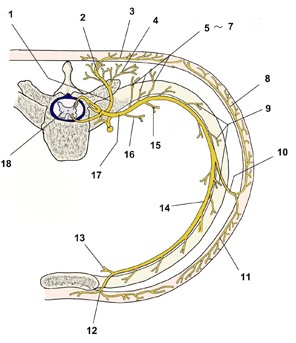 Thoracic spinal nerve and intercostal nerve branches (transverse schematic)
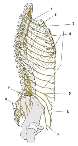 Intercostal nerves and nerves of the trunk wall (lateral view)
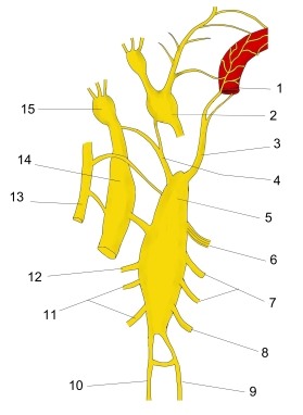
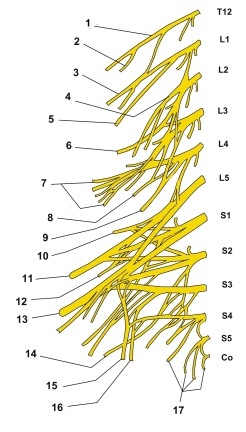 Lumbosacral plexus (schematic)
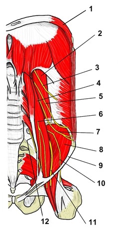 Lumbar plexus on the posterior abdominal wall (anterior view)
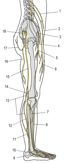 Lumbar and sacral plexuses, right lower limb (lateral view)
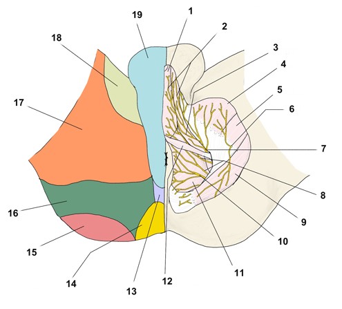 Pudendal plexus: nerves and cutaneous territories of the male perineum (inferior view)
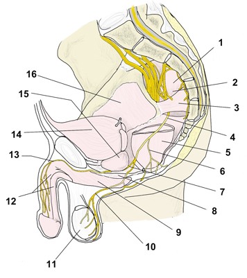 Pudendal plexus (median section)
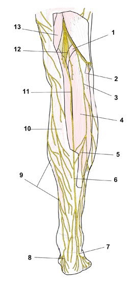 Nerves of the leg (posterior view)
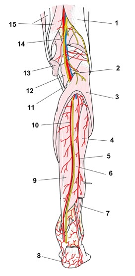 Leg, nerves and arteries (posterior view)
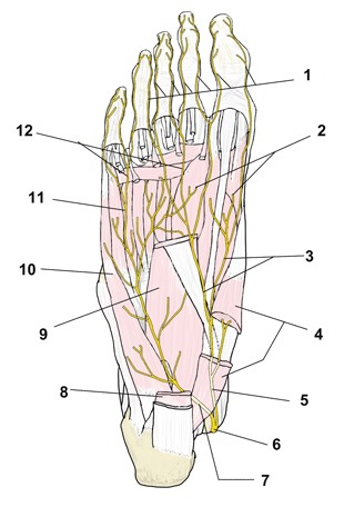 Nerves of the sole of the foot
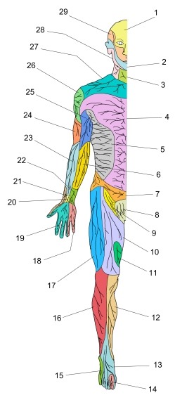 Cutaneous nerve distribution (anterior view)
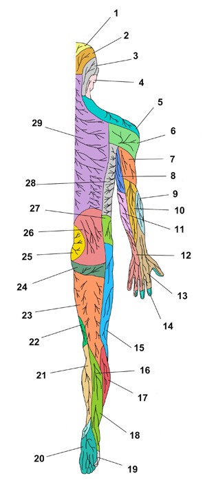 Cutaneous nerve distribution (posterior view)