← Multi-nerve illustrations: Cranial nerves
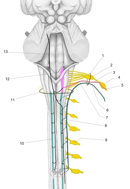
| No. | Name | Page |
|---|---|---|
| 1 | Jugular foramen | Detail |
| 2 | Superior ganglion (vagus nerve) | Detail |
| 3 | Internal branch (accessory nerve) | — |
| 4 | Inferior ganglion (vagus nerve) | Detail |
| 5 | Fibers of internal branch to laryngeal muscles (via recurrent laryngeal nerve) | — |
| 6 | External branch (accessory nerve) | — |
| 7 | Cranial root (accessory nerve) | — |
| 8 | Spinal root (accessory nerve) | — |
| 9 | Spinal ganglion | Detail |
| 10 | Spinal nucleus of accessory nerve | — |
| 11 | Foramen magnum | Detail |
| 12 | Nucleus ambiguus | — |
| 13 | Corticonuclear fibers | — |
More illustrations: Cranial nerves
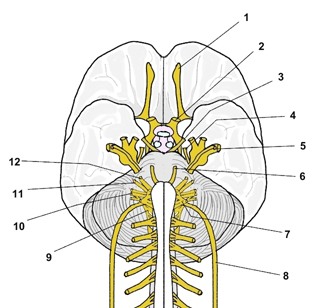 Cranial nerves I to XII on the base of the brain (schematic)
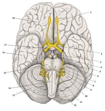 Cranial nerves on the base of the brain (inferior view, overview)
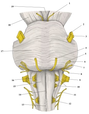 Cranial nerves at the brainstem (anterior view, enlarged)
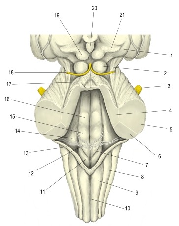 Brainstem with cerebellum removed (posterior view)
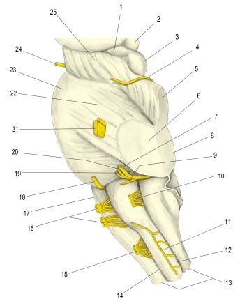 Cranial nerves and brainstem, cerebellum removed (left lateral view)
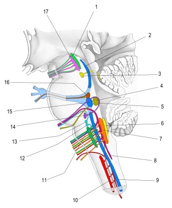 Cranial nerve nuclei in the brainstem (lateral view)
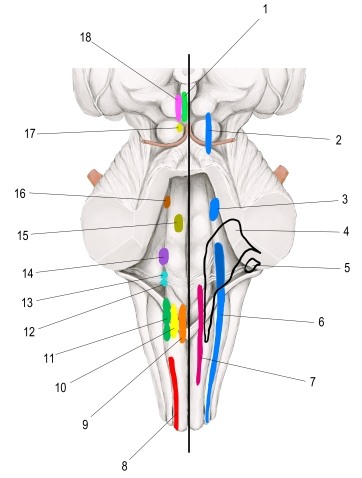 Cranial nerve nuclei (projection on brainstem)
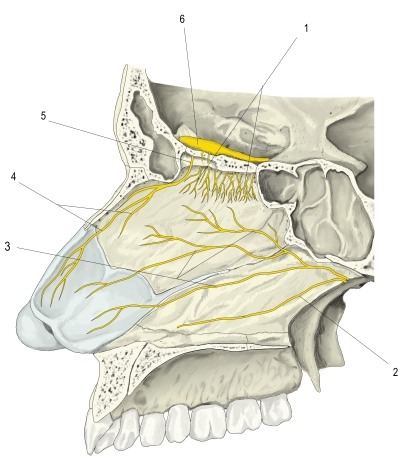 Nerves of the nasal septum (left lateral view)
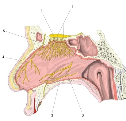 Nerves of the nasal septum, mucosa (left lateral view)
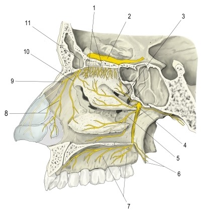 Nerves of the lateral wall of the right nasal cavity
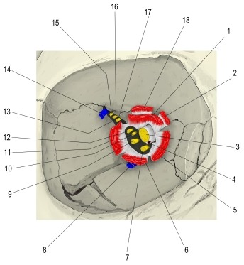 Superior orbital fissure, left (anterior view)
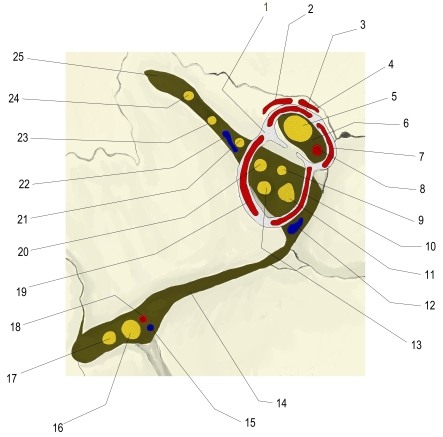 Orbital fissures and common tendinous ring (anterior view)
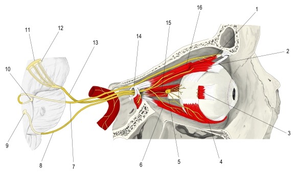 Oculomotor nerve and nuclei, right orbit (lateral view)
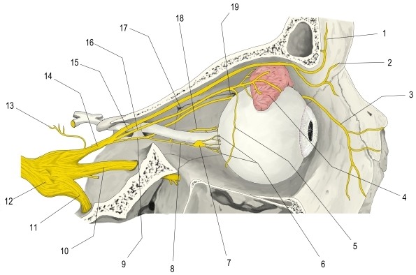 Ophthalmic nerve in the orbit (lateral view, sagittal section)
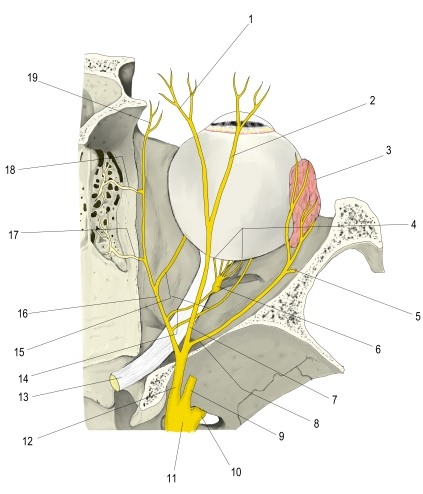 Ophthalmic nerve in the right orbit (superior view, horizontal section)
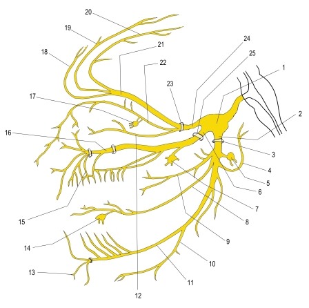 Trigeminal nerve and its three divisions (schematic)
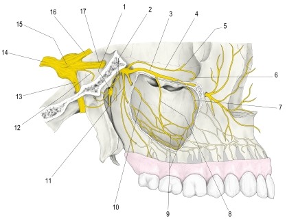 Maxillary nerve (lateral view)
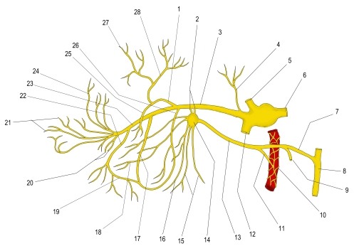 Maxillary nerve, branches (schematic)
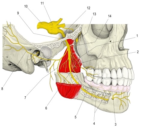 Mandibular nerve (lateral view)
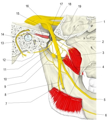 Mandibular nerve and otic ganglion (medial view)
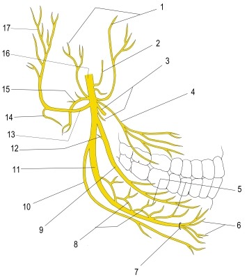 Mandibular nerve, branches (schematic)
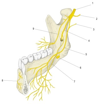 Mandibular nerve and its branches
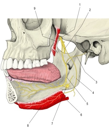 Lingual nerve and submandibular ganglion (lateral view, mandible removed)
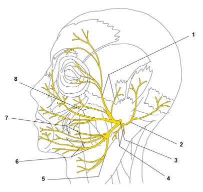 Facial nerve, branches in the face (lateral view)
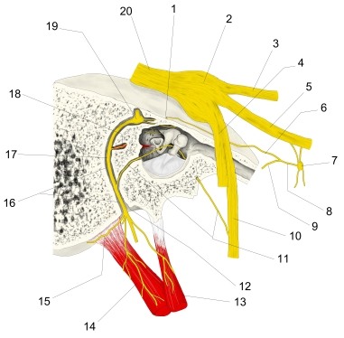 Facial nerve in the petrous part of the right temporal bone (lateral view)
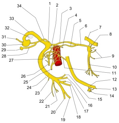 Facial nerve (schematic)
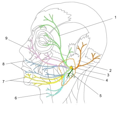 Facial nerve, branches after the stylomastoid foramen
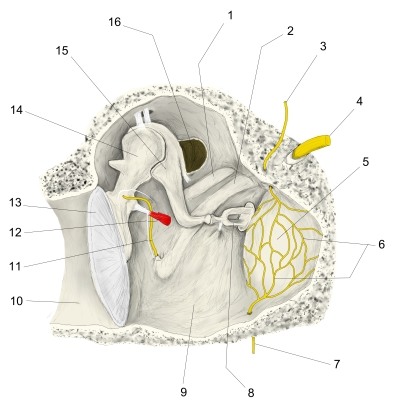 Walls of tympanic cavity (anterior wall removed)
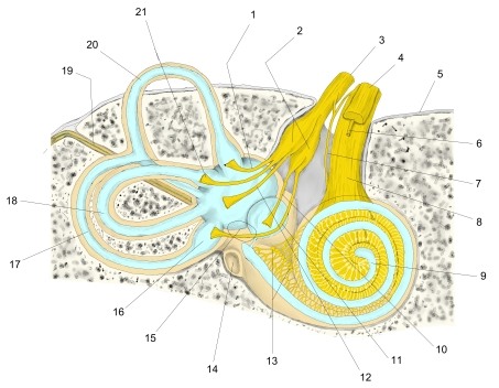 Vestibulocochlear nerve and inner ear in the temporal bone, right ear (anterior view)
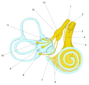 Vestibulocochlear nerve and membranous labyrinth, right ear (anterior view, simplified)
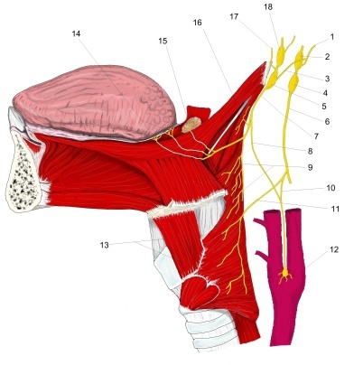 Glossopharyngeal nerve (schematic)
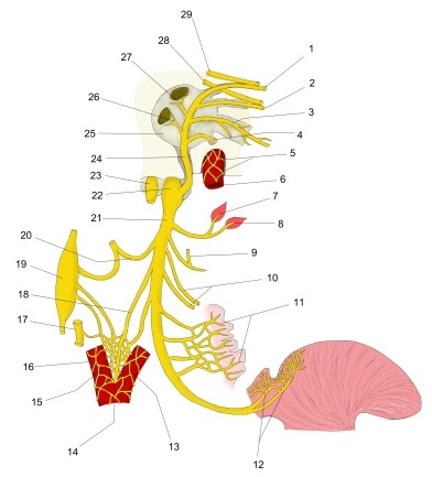 Right glossopharyngeal nerve (schematic)
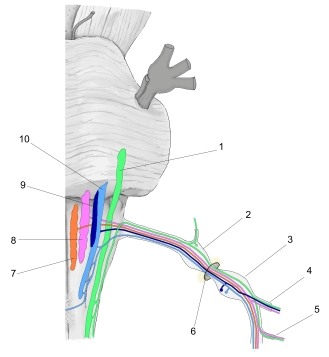 Vagus nerve, nuclei of origin (anterior view, schematic)
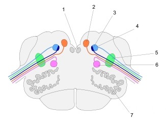 Vagus nerve, nuclei of origin (section at level of olive)
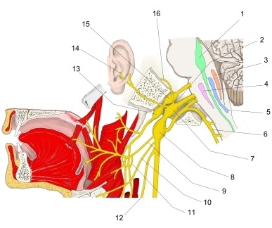 Vagus nerve, head and neck, with brainstem nuclei
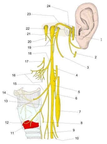 Vagus nerve, head and neck (schematic)
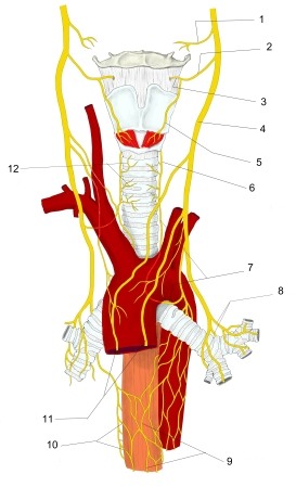 Vagus nerve, neck and thorax (anterior view, schematic)
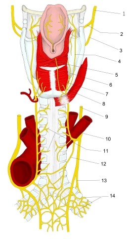 Vagus nerve, head and neck (schematic, posterior view)
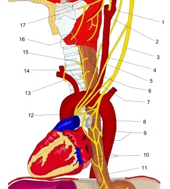 Vagus nerve, neck and thorax, branches (schematic)
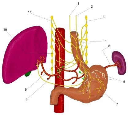 Vagus nerve, thorax and abdomen (schematic)
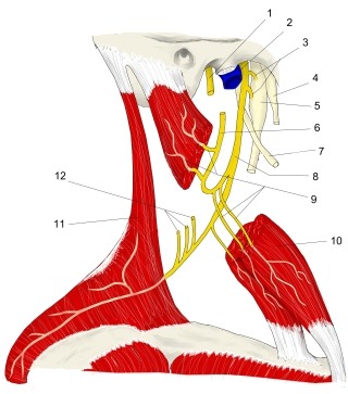 Accessory nerve, external branch (schematic, right lateral view)
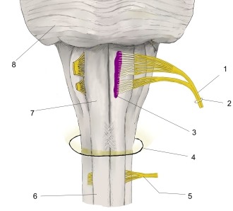 Hypoglossal nerve and its nucleus (schematic)
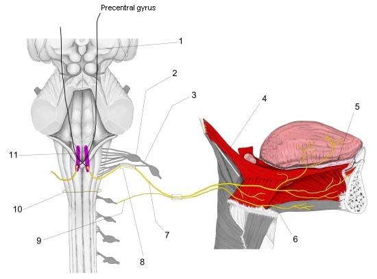 Hypoglossal nerve, nucleus and course (schematic I)
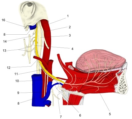 Hypoglossal nerve (schematic II)