Latin: rami alveolares superiores posteriores
Overview
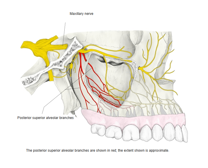
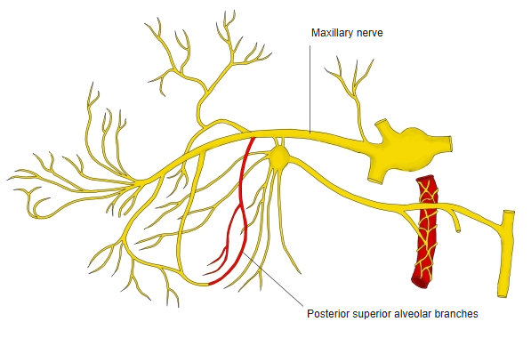
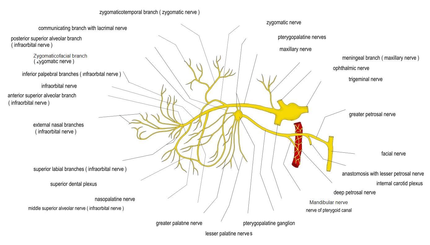
- Branches of the maxillary nerve in the pterygopalatine fossa, before it enters the orbit (some texts count them as branches of the infraorbital nerve).
- Descend on the maxillary tuberosity with the posterior superior alveolar artery.
Posterior superior alveolar branches 
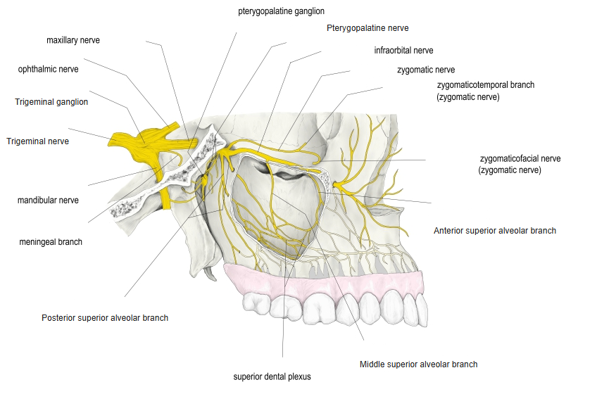
Maxillary nerve, lateral view
Distribution
| Part | Supply |
|---|---|
| Branches staying outside the bone | Gums of the molar region and the adjacent cheek mucosa |
| Branches through the alveolar foramina | Lining of the maxillary sinus and the upper molar teeth (with the middle superior alveolar branch, they form the superior dental plexus) |
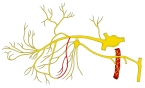
Posterior superior alveolar branches (schematic) 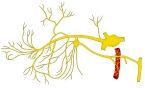
Branches of the maxillary nerve 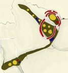
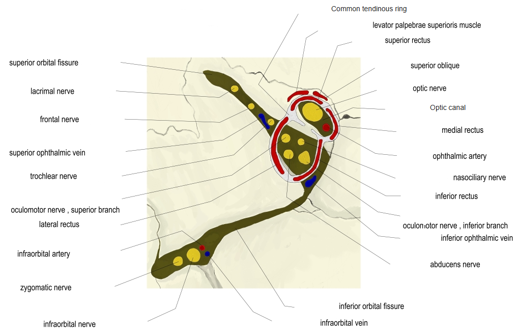
Orbital fissures and optic canal
References and sources
- Further reading: Posterior superior branches, Wikipedia.