Latin: nervus maxillaris
Overview
- The second division of the trigeminal nerve (V2); sensory.
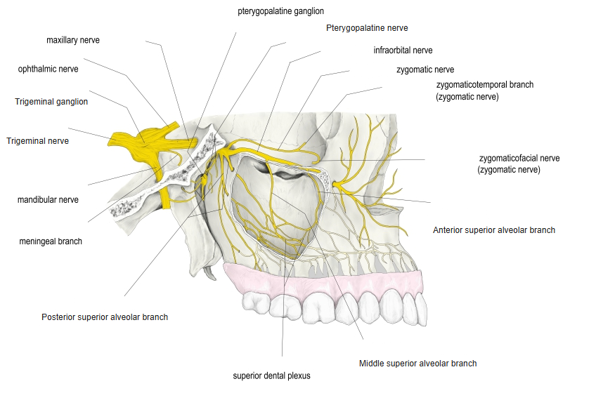
- Leaves the skull through the foramen rotundum into the pterygopalatine fossa, where it gives the zygomatic nerve and the pterygopalatine nerves.
- Continues as the infraorbital nerve through the inferior orbital fissure into the orbit.
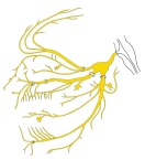
Trigeminal nerve and its branches (schematic)
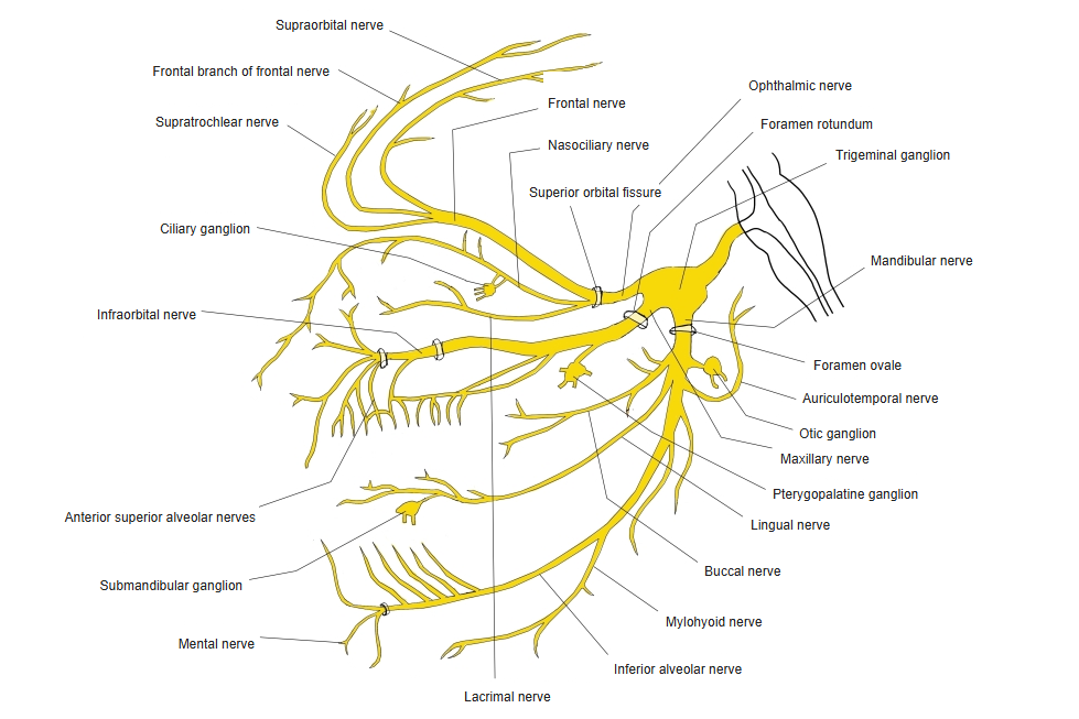
Branches
| Branch | Supply |
|---|---|
| Meningeal branch | Dura of the middle cranial fossa |
| Zygomatic nerve | Skin of the temple and cheek; secretory fibres to the lacrimal nerve |
| Pterygopalatine nerves | To the pterygopalatine ganglion: nose, palate, pharynx |
| Posterior superior alveolar branches | Molars, gums, maxillary sinus |
| Infraorbital nerve | Upper teeth (middle and anterior superior alveolar branches), lower eyelid, side of the nose, upper lip |
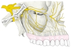
Maxillary nerve, lateral view 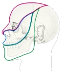
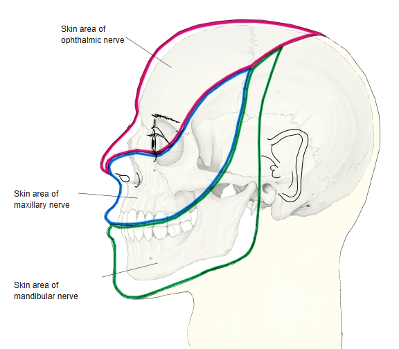
Skin areas of the trigeminal nerve
References and sources
- Further reading: Maxillary nerve, Wikipedia.