Latin: macula lutea
Overview
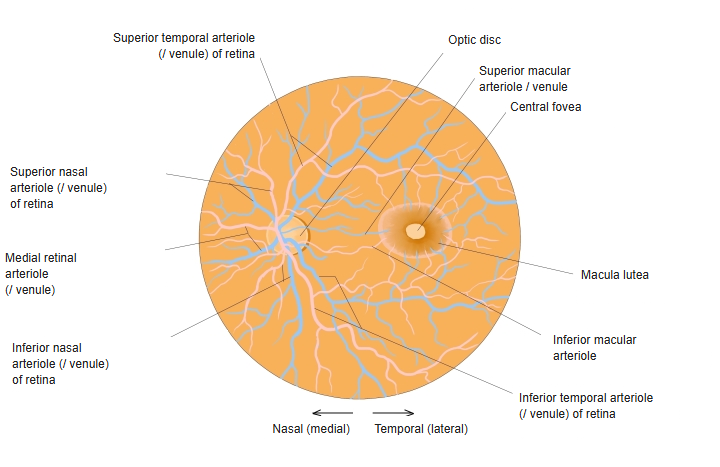
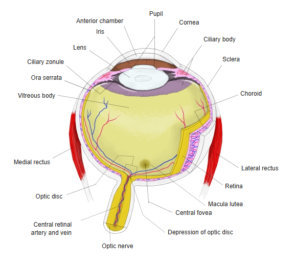
- A small yellowish area of the retina at the posterior pole, lateral (temporal) to the optic disc.
- The yellow colour comes from carotenoid pigments (lutein and zeaxanthin) that filter blue light.
- About 5 mm across anatomically; clinically the central part, about 1.5 mm, is often called the macula.
- In its centre is the central fovea, the region of the sharpest and colour vision.

Optic disc and macula in the fundus 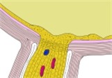
Optic disc and macula, section
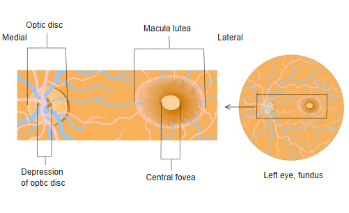
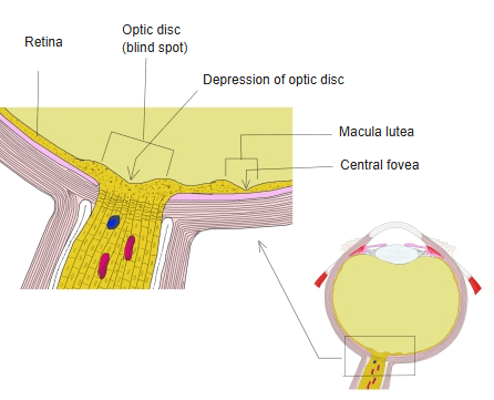
Central fovea and its zones
| Zone (centre outward) | Features |
|---|---|
| Foveola | The floor of the pit, about 0.35 mm; only cones, very densely packed |
| Foveal avascular zone | Central area without retinal capillaries |
| Fovea | The pit, about 1.5 mm across; the inner retinal layers are pushed aside so that light reaches the cones directly |
| Parafovea and perifovea | Surrounding rings where the displaced ganglion cells and inner nuclear cells pile up |
- The fovea has no rods, so it is relatively blind in very dim light.
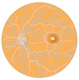
Fundus of the eye 
Eyeball, horizontal section
References and sources
- Further reading: Macula lutea, Wikipedia.