Thyrohyoid membrane (Latin: membrana thyrohyoidea)

Also called the hyothyroid membrane

The thyrohyoid membrane is a fibroelastic membrane stretched between the hyoid bone and the upper border of the thyroid cartilage, suspending the larynx from the hyoid.
It forms most of the front wall of the larynx above the thyroid cartilage

· Below, it is attached to the upper border of the thyroid cartilage and the front of its superior horns; above, to the upper margin of the back of the body of the hyoid and to the greater horns.

· It passes up behind the body of the hyoid, separated from it by a small bursa that lets the larynx glide upwards in swallowing.

· In front of it lie the thyrohyoid, sternohyoid and omohyoid, three of the infrahyoid muscles.

Some textbooks count the thyrohyoid membrane among the ligaments of the larynx (as an extrinsic membrane; one Japanese textbook lists it as one of eight), while others treat it with the hyoid bone. Sources differ.
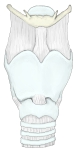
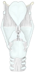
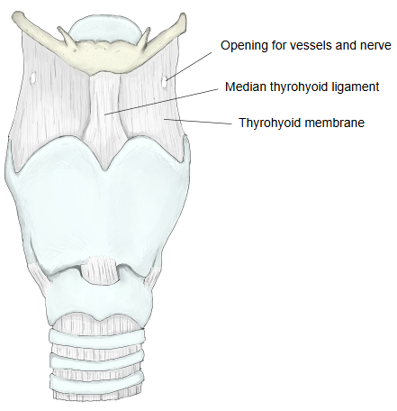
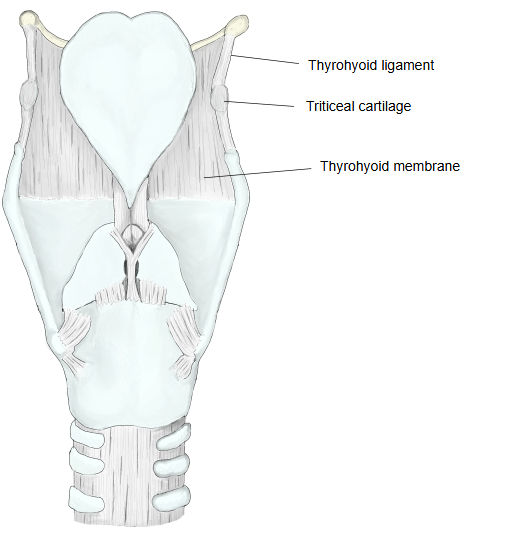
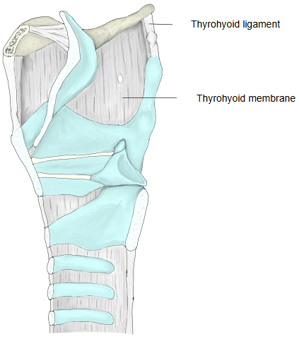
The middle part and the two back edges of the thyrohyoid membrane are thickened; they are called the median thyrohyoid ligament and the thyrohyoid ligament.
Median thyrohyoid ligament: the thick middle part, in frontThyrohyoid ligament: the thick free back edge on each side; it often contains a small triticeal cartilage
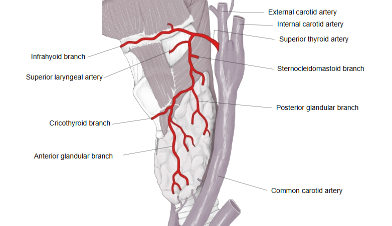
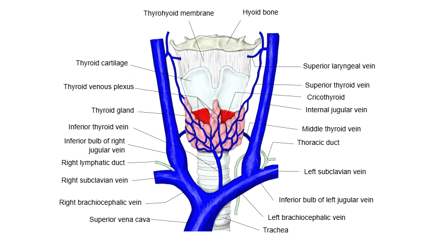
On each side the thyrohyoid membrane has a small opening for vessels and a nerve: the superior laryngeal artery, the superior laryngeal vein and the internal branch of the superior laryngeal nerve.
The artery and vein serve the inside of the larynx; the nerve branch carries sensation from the larynx above the vocal folds
  • The larynx and hyoid bone from the front, showing the thyrohyoid membrane, the median thyrohyoid ligament and the opening for the superior laryngeal vessels and nerve.Larynx (anterior)
  • The larynx from behind, showing the thyrohyoid membrane, the thyrohyoid ligament and the triticeal cartilage.Larynx (posterior)
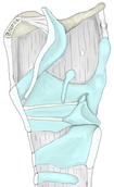
  • A median section of the larynx seen from the inside, showing the thyrohyoid membrane and the thyrohyoid ligament.Larynx (median section)
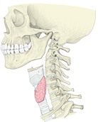
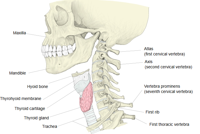
  • The skull, cervical spine and larynx from the side, showing where the thyrohyoid membrane lies between the hyoid bone and the thyroid cartilage.Head and neck (lateral)
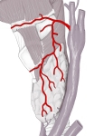
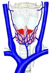
  • The superior thyroid artery and its branches, including the superior laryngeal artery, which pierces the thyrohyoid membrane.Superior thyroid artery
  • The veins in front of the larynx and thyroid gland, including the superior laryngeal vein near the thyrohyoid membrane.Veins of the thyroid region

Reference websites

[Wikipedia]

· Thyrohyoid membrane, Wikipedia.

[Websites with illustrations]

· Larynx, external view (labelled) (Wikimedia Commons)

· Larynx: cartilages, ligaments and muscles (Kenhub)

· The larynx (TeachMeAnatomy)

Where it is found

References and sources

  1. Further reading: Thyrohyoid membrane, Wikipedia.
  2. Further reading: Larynx, Wikipedia.
  3. Federative International Programme for Anatomical Terminology (FIPAT). Terminologia Anatomica, 2nd ed. (TA2), 2019.