Larynx (Latin: larynx)
- Overview
- Laryngeal cartilages
- Joints and ligaments
- Muscles (intrinsic, extrinsic)
- Mucosal folds
- Divisions of the laryngeal cavity
- Structure (mucosa, submucosa)
- Vessels and nerves
- Development
- Related terms
- References
Overview
system
- Airway
- Nasal cavity : from the nostrils to the choanae; divided into right and left halves by the nasal septum and lined with mucosa.
- Pharynx : made of three parts (upper, middle, lower) and lined with mucosa; also part of the digestive system.
- Larynx : funnel-shaped tube at the level of the 4th–7th cervical vertebrae; its framework is cartilage.
- Trachea : tube about 10 cm long and 20–25 mm wide; divides at the level of the 4th–5th thoracic vertebrae.
- Bronchi : branch from the trachea to the right and left, and divide into many bronchial branches inside the lungs.
- Lungs : right lung (upper, middle, lower lobes), left lung (upper, lower lobes); gas exchange (external respiration) takes place in the alveoli.
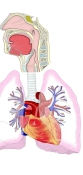
Note: the height of the larynx changes with age. In the newborn it lies two or three vertebrae higher than in the adult, which lets the infant breathe and swallow at the same time; it reaches its adult position after about four years of age, and in old age it sits lower still.
In front, the middle of the larynx is covered only by the superficial cervical fascia, while its front and sides are covered by the platysma, the infrahyoid muscles and the thyroid gland.
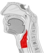 Larynx (median section)
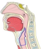 Larynx (median section, 2)
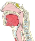 Nasal cavity to larynx (median section)
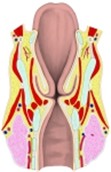 Larynx (coronal section)
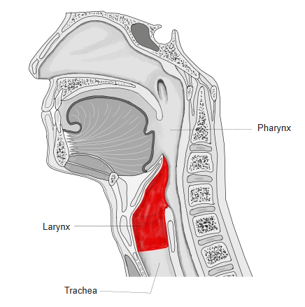
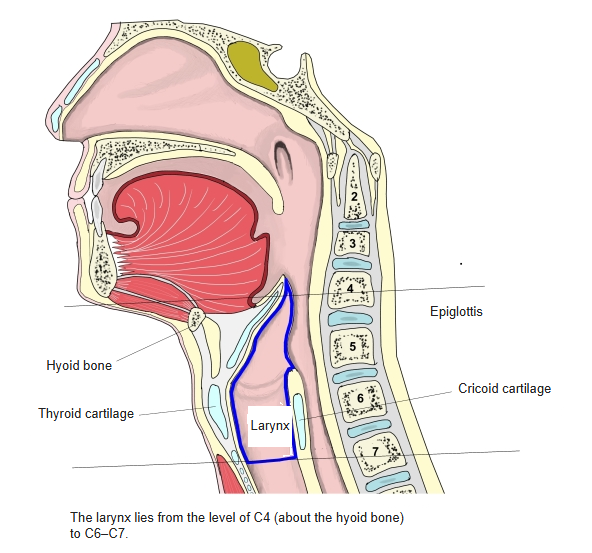
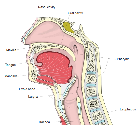
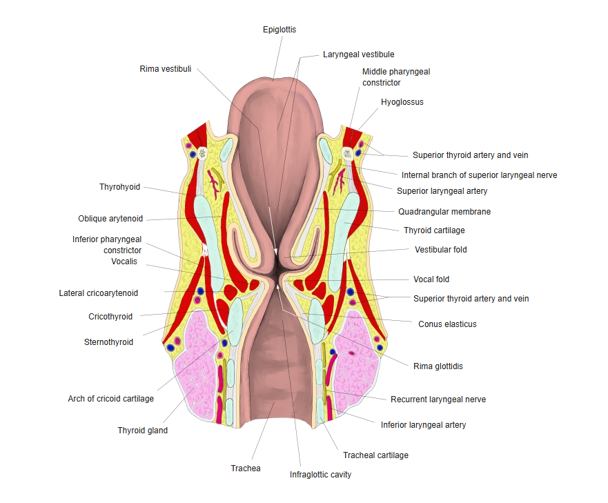
Laryngeal cartilages
The cartilages of the larynx form its essential framework. They fall into two groups, unpaired and paired. See the page of each cartilage for details.
Most laryngeal cartilages are hyaline cartilage; the epiglottic cartilage, the vocal process of the arytenoid, and the corniculate and cuneiform cartilages are elastic cartilage. The thyroid cartilage, and then the cricoid cartilage, begin to ossify relatively early.
cartilages
- Unpaired
- Thyroid cartilage : the largest laryngeal cartilage; forms most of the front wall of the larynx; laryngeal prominence.
- Cricoid cartilage : the lowest laryngeal cartilage; joined to the tracheal cartilage by the cricotracheal ligament.
- Epiglottic cartilage : the framework of the epiglottis.
- Paired
- Arytenoid cartilage : sits on the lamina of the cricoid cartilage; the most important cartilage for voice production.
- Corniculate cartilage : a very small cartilage on the apex of the arytenoid cartilage.
- Cuneiform cartilage : a small, flat cartilage in front of and lateral to the corniculate cartilage; may be absent.
- Sesamoid cartilages : small cartilages of variable number.
Cartilages with an orange border are hyaline; the others are elastic. Some sources do not count the corniculate and cuneiform cartilages among the laryngeal cartilages.
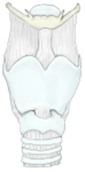 Laryngeal cartilages
(anterior view)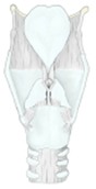 Laryngeal cartilages
(posterior view)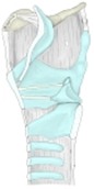 Laryngeal cartilages
(median section)
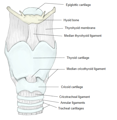
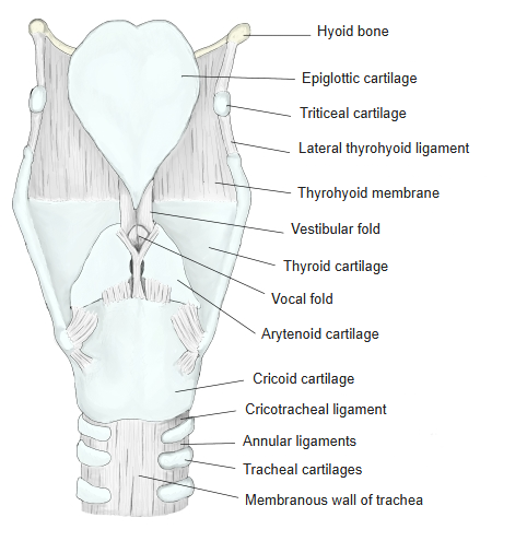
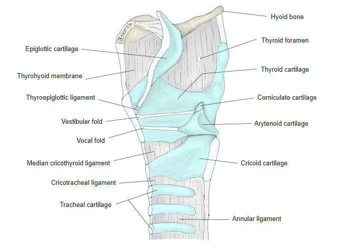
Joints and ligaments
[Joints]
The larynx has the following two joints.
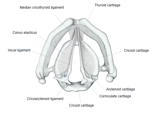
1. Cricothyroid joint: between the thyroid articular surface of the cricoid cartilage and the inferior horn of the thyroid cartilage; reinforced by the capsule of the cricothyroid joint.
2. Cricoarytenoid joint: between the arytenoid articular surface of the cricoid cartilage and the base of the arytenoid cartilage. Its capsule is reinforced by the posterior cricoarytenoid ligament, a thickening of its back part.
[Ligaments]
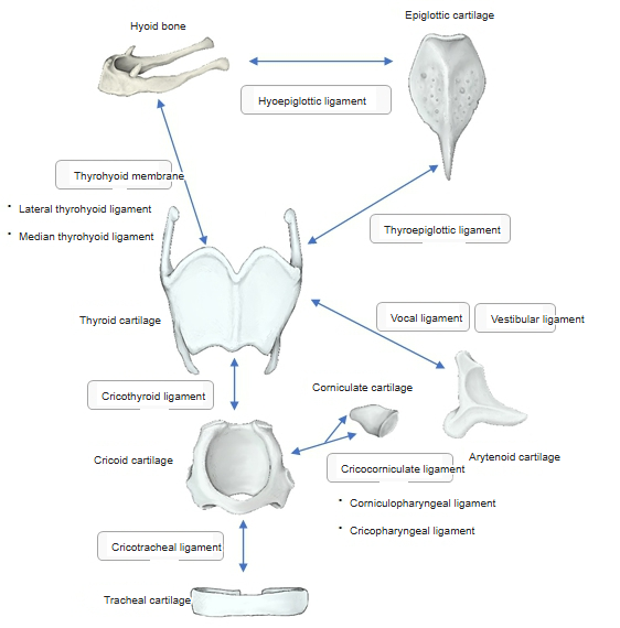
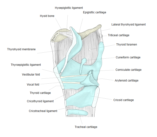
The ligaments of the larynx are listed below.
| 1 | Thyrohyoid membrane | A membrane of elastic connective tissue between the lower border of the hyoid bone and the upper border of the thyroid cartilage. Its thicker parts are called the median thyrohyoid ligament and the lateral thyrohyoid ligament. |
| 2 | Hyoepiglottic ligament | Runs from the upper border of the body of the hyoid bone to the front (lingual) surface of the epiglottic cartilage. |
| 3 | Thyroepiglottic ligament | Joins the stalk of the epiglottis to the thyroid cartilage. |
| 4 | Cricothyroid ligament | Joins the arch of the cricoid cartilage to the lower border (inferior thyroid notch) of the thyroid cartilage. Its parts are called the median cricothyroid ligament and the lateral cricothyroid ligament. |
| 5 | Cricotracheal ligament | Joins the lower border of the cricoid cartilage to the first tracheal cartilage. |
| 6 | Cricocorniculate ligament | Y-shaped, with three limbs: two attach to the corniculate cartilages and one to the cricoid cartilage. The upper limbs are also called the cricocorniculate (corniculopharyngeal) parts, and the lower limb the cricopharyngeal ligament. |
| 7 | Vestibular ligament | Stretches between the inner surface of the angle of the thyroid cartilage and the arytenoid cartilage (near its colliculus). |
| 8 | Vocal ligament | Stretches from the inner surface of the thyroid cartilage, next to the midline, to the vocal process of the arytenoid cartilage. |
The vestibular and vocal ligaments are the free borders of the quadrangular membrane and the conus elasticus, the two parts of the fibroelastic membrane of the larynx.
 Laryngeal cartilages and ligaments
- Laryngeal cartilages
(anterior view) - Laryngeal cartilages
(posterior view) 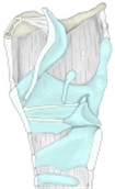 Laryngeal cartilages
(median section)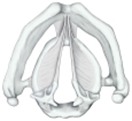 Laryngeal cartilages
(superior view)
Muscles
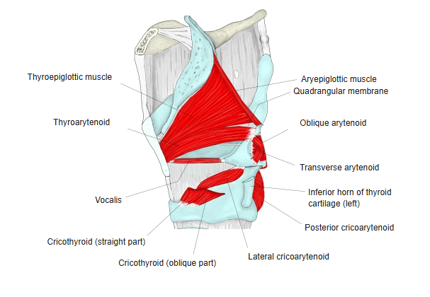
The larynx is moved by many small muscles. They are divided into extrinsic and intrinsic laryngeal muscles.
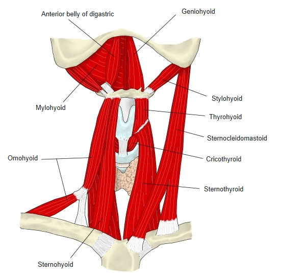
· Extrinsic laryngeal muscles: change the position of the cartilages (the larynx as a whole).
· Intrinsic laryngeal muscles: change the shape of the glottis and the tension of the vocal folds.
[Intrinsic laryngeal muscles]
The chart below lists the intrinsic laryngeal muscles.
laryngeal
muscles
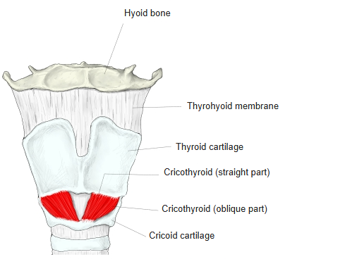
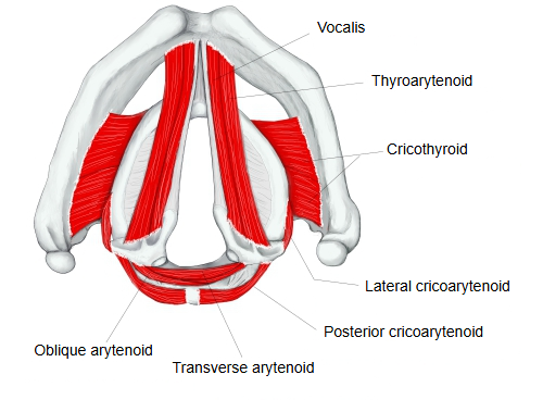
- Cricothyroid : origin) outer lower border of the arch of the cricoid cartilage; insertion) lower border and inferior horn of the thyroid cartilage.
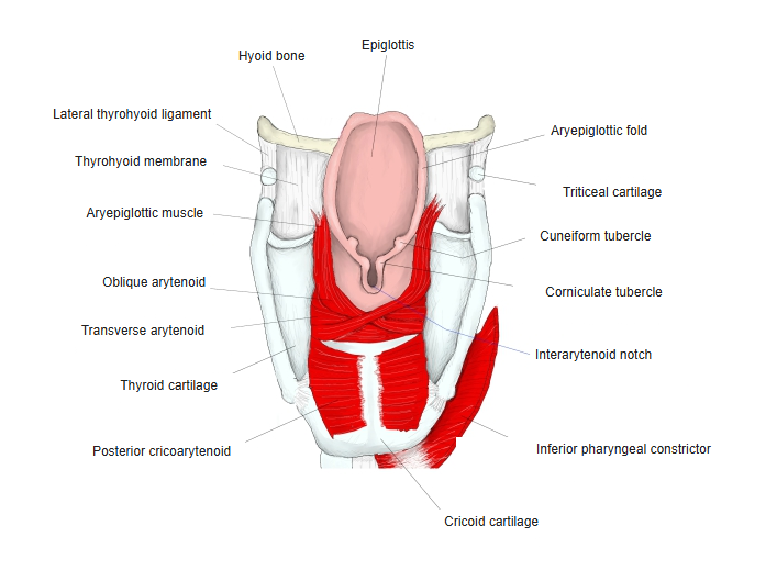
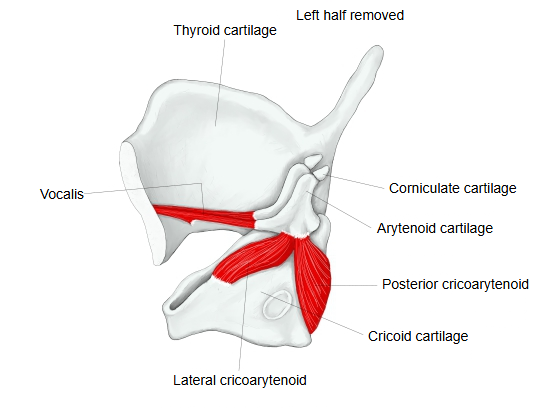
- Posterior cricoarytenoid : origin) hollow on the back of the lamina of the cricoid cartilage; insertion) muscular process of the arytenoid cartilage.
- Lateral cricoarytenoid : origin) upper border and outer surface of the side of the cricoid arch; insertion) muscular process and front of the arytenoid cartilage.
- Arytenoid : fills the hollows on the backs of both arytenoid cartilages and inserts on their lateral borders. Two parts:
- Transverse arytenoid : joins the lateral borders of the two arytenoid cartilages horizontally.
- Oblique arytenoid : origin) muscular process of one arytenoid cartilage; insertion) apex of the other.
- Aryepiglotticus : continuation of the oblique arytenoid from the apex of the arytenoid cartilage into the aryepiglottic fold, to the epiglottic cartilage.
- Thyroarytenoid : origin) inner surface of the thyroid cartilage; insertion) muscular process and oblong fovea of the arytenoid cartilage. Some fibres continue to the epiglottis as the thyroepiglotticus. Inside the laryngeal cavity it is divided into:
- Vocalis : origin) back of the thyroid cartilage next to the midline; insertion) vocal process and oblong fovea of the arytenoid cartilage.
- Ventricularis : origin) back of the thyroid cartilage next to the midline; insertion) near the colliculus of the arytenoid cartilage.
- Oblique thyroarytenoid : origin) muscular process of the arytenoid cartilage; insertion) upper border of the thyroid cartilage (inconstant).
Sources group the intrinsic muscles differently. Many list six: cricothyroid, posterior cricoarytenoid, lateral cricoarytenoid, transverse and oblique arytenoid, and thyroarytenoid (with the vocalis); others treat the vocalis, the ventricularis and the oblique thyroarytenoid as parts of the thyroarytenoid.
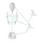 Laryngeal cartilages and muscles
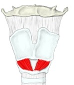 Larynx (anterior view)
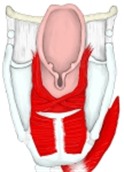 Larynx (posterior view)
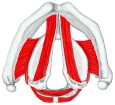 Larynx (superior view)
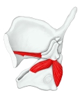 Larynx (right half, medial view)
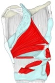 Intrinsic laryngeal muscles
(median section)
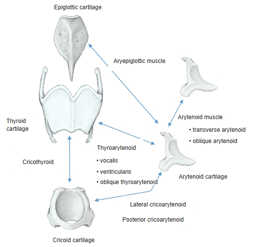
[Extrinsic laryngeal muscles]
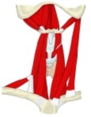
The extrinsic laryngeal muscles attach the larynx to its surroundings and raise or lower it as a whole, for example in swallowing. They include the infrahyoid muscles that attach to the thyroid cartilage (the sternothyroid and thyrohyoid), and pharyngeal muscles that attach to the laryngeal cartilages (the inferior pharyngeal constrictor, stylopharyngeus and palatopharyngeus). The suprahyoid muscles also move the larynx indirectly through the hyoid bone.
 Muscles from the manubrium to the mandible
Mucosal folds
The laryngeal mucosa, like that of the trachea and bronchi, is lined with ciliated epithelium (pseudostratified ciliated columnar epithelium); only the surface of the vocal folds is covered, like the skin, with stratified squamous epithelium.
recesses of
the larynx
- In the cavity
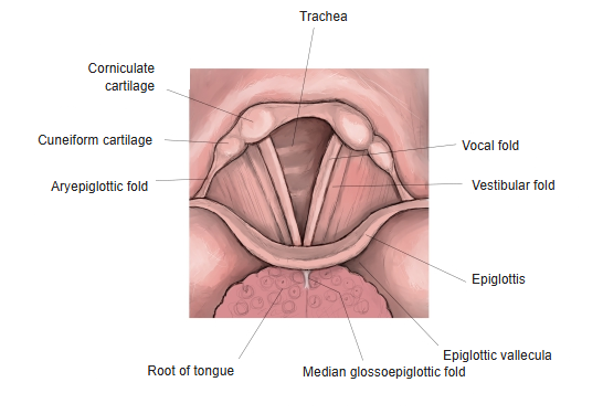
- Vestibular fold : fold over the vestibular ligament and the quadrangular membrane (= ventricular fold, false vocal cord).
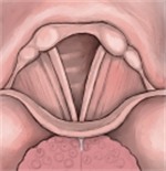
- Vocal fold : fold over the vocal ligament and the conus elasticus; ⇒ responsible for "voice production".
- At the inlet
- Aryepiglottic fold : forms both sides of the laryngeal inlet.
- In front
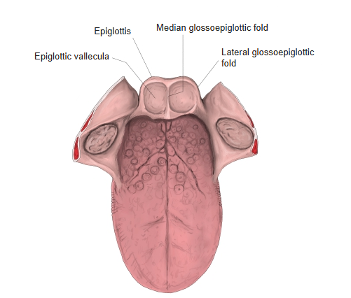
- Median glossoepiglottic fold : between the midline of the tongue and the epiglottic cartilage.
- Lateral glossoepiglottic fold : runs on each side of the median fold, across the epiglottic vallecula.
- Others
- Fold of superior laryngeal nerve : a fold in the wall of the piriform recess where the superior laryngeal nerve runs.
- Pharyngoepiglottic fold : from the side wall of the pharynx to the side of the epiglottic cartilage.
- Larynx (coronal section)
 Vocal folds
- Larynx (posterior view)
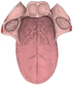 Root of the tongue
Divisions of the laryngeal cavity
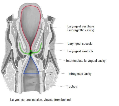
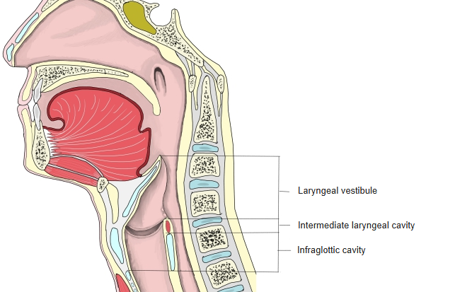
The laryngeal cavity is divided into three parts: the vestibule above, which begins at the inlet; the middle part, which holds the glottis, essential for the voice; and the infraglottic cavity below, which continues into the trachea.
the laryngeal
cavity
- Laryngeal vestibule : upper part, from the laryngeal inlet to the vestibular folds = supraglottic cavity.
- Intermediate laryngeal cavity : between the vestibular and vocal folds; bounded above by the rima vestibuli and below by the rima glottidis ⇒ contains the glottis.
- Laryngeal ventricle : the space bulging outwards on each side of the cavity; its upward extension at the front is the laryngeal saccule.
- Infraglottic cavity : from the rima glottidis down to the trachea (lower border of the cricoid cartilage, level of the 6th cervical vertebra).
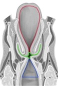 Divisions of the cavity
(coronal section)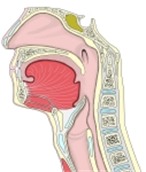 Divisions of the cavity
(median section)
Structure
The wall of the larynx is made of the following layers.
| 1 | Mucosa | Epithelium | Pseudostratified ciliated columnar epithelium: lines most of the larynx, like the nasal mucosa. The cilia beat towards the mouth. Stratified squamous epithelium: covers the vocal folds, the edge of the laryngeal inlet (aryepiglottic folds) and the front surface and upper back surface of the epiglottis. |
| Lamina propria | Rich in elastic fibres. Lymphocytes, mast cells, eosinophils and plasma cells are scattered in it, with lymphoid nodules here and there (those in the laryngeal ventricle are the laryngeal lymphoid nodules). | ||
| 2 | Submucosa | Rich in membranous elastic fibres, forming the fibroelastic membrane of the larynx. It contains many glands, together called the laryngeal glands (mixed tubuloacinar glands), named by position: 1) anterior laryngeal glands, on the laryngeal surface of the epiglottic cartilage; 2) middle laryngeal glands, in the vestibular folds; 3) posterior laryngeal glands, between the arytenoid and corniculate cartilages. | |
Vessels and nerves
The vessels and nerves of the larynx are as follows.
| Arteries | Superior thyroid artery (from the external carotid artery) | Gives two branches: · Superior laryngeal artery: enters the larynx through the thyrohyoid membrane. · Cricothyroid branch: reaches the cricothyroid muscle and enters the larynx through the cricothyroid ligament. | 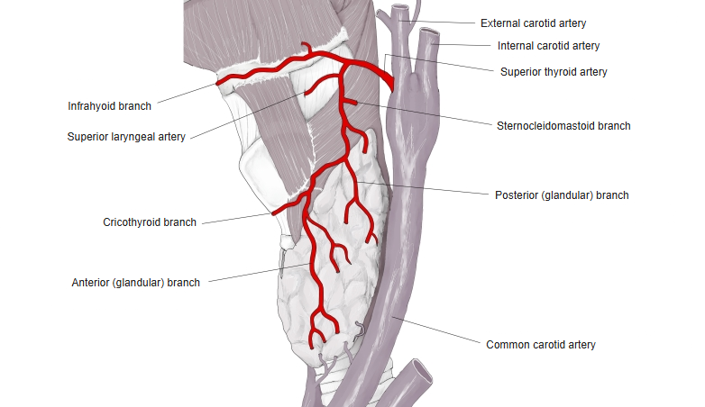 |
| Inferior thyroid artery (from the thyrocervical trunk of the subclavian artery) | The inferior laryngeal artery, a branch of the inferior thyroid artery, enters from below and supplies the muscles and mucosa of the larynx. | 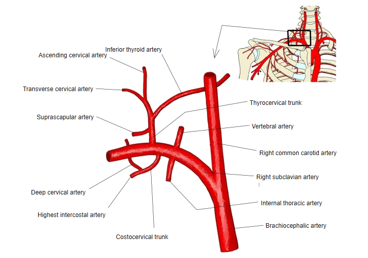 | |
| Veins | Run with the arteries. The superior laryngeal vein drains into the internal jugular vein (through the superior thyroid vein), and the inferior laryngeal vein into the brachiocephalic vein (through the inferior thyroid vein). | 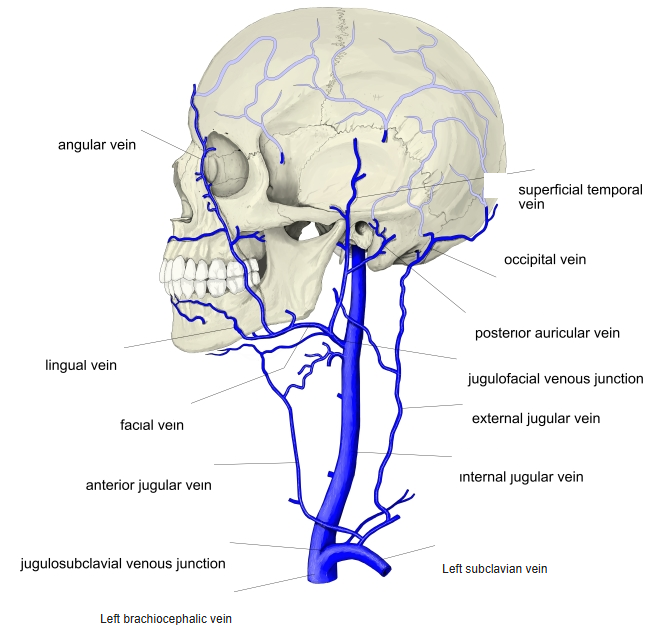 | |
| Lymphatics | The upper part of the larynx drains to the infrahyoid nodes and on to the deep cervical lymph nodes; the lower part to the pretracheal and paratracheal nodes. The regions above and below the vocal folds are fairly separate, except at the back. Lymph from the piriform recess and the epiglottis goes to the nodes beside the thyroid gland. | ||
| Nerves | Vagus nerve | The following branches supply the larynx: · Superior laryngeal nerve. Internal branch: sensory; passes through the thyrohyoid membrane into the larynx and supplies the mucosa above the glottis. External branch: motor; runs outside the inferior pharyngeal constrictor to the cricothyroid muscle. · Inferior laryngeal nerve (end branch of the recurrent laryngeal nerve; mixed). Sensory fibres: mucosa below the glottis; they also join branches of the superior laryngeal nerve. Motor fibres: all laryngeal muscles except the cricothyroid. Small branches from the sympathetic trunk also join the plexus formed by the vagal branches in the larynx. | 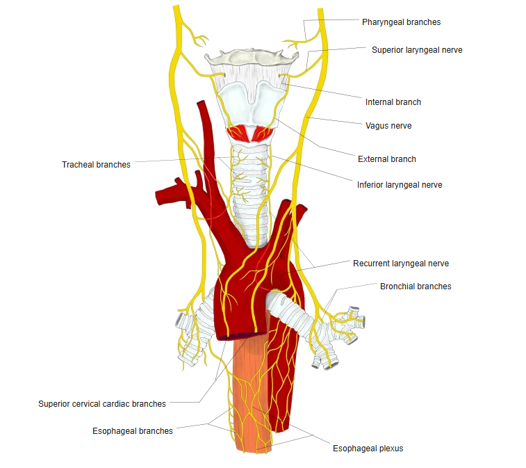 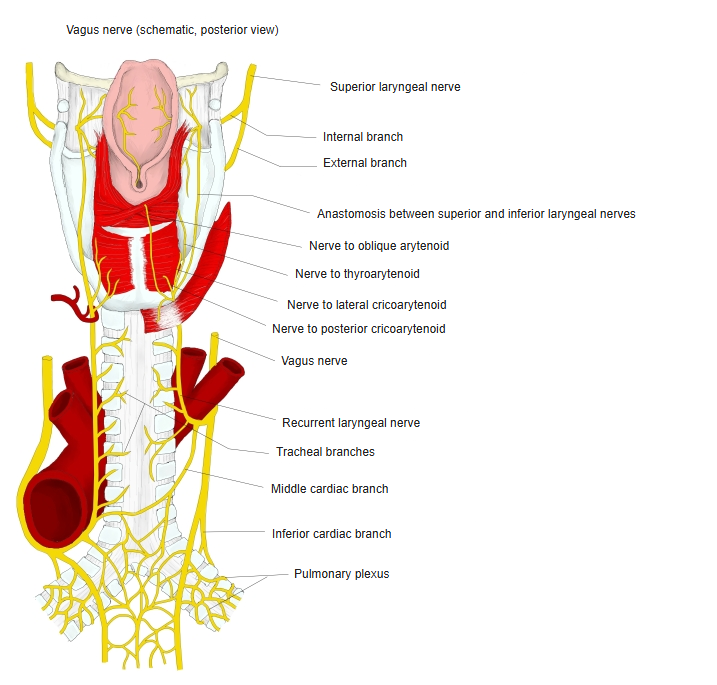 |
Development
The larynx develops, together with the trachea and lungs, from the respiratory diverticulum that grows from the front wall of the foregut. Its epithelium and glands come from the endoderm of the foregut, while its cartilages, muscles, vessels and nerves come from the mesenchyme of the pharyngeal arches around the opening of the diverticulum.
A pair of swellings (the arytenoid swellings) forms on each side of the opening; the arytenoid cartilages develop inside them, and the epiglottis develops from a swelling in front of them. The epithelium of the larynx grows so fast that it closes the lumen for a time; when the lumen opens again, the laryngeal ventricles appear on each side, bounded above by the vestibular folds and below by the vocal folds.
The thyroid cartilage comes from the fourth pharyngeal arch, and the cricoid and arytenoid cartilages from the sixth. Accordingly, the superior laryngeal nerve is the nerve of the fourth arch and the recurrent laryngeal nerve that of the sixth.
Related terms
Main anatomical terms related to the larynx (other than muscles, nerves, vessels, cartilages and their parts), in alphabetical order.
| A | Anterior laryngeal glands, Anterior macula flava, Aryepiglottic fold |
| C | Conus elasticus, Corniculate tubercle, Cricoarytenoid joint, Cricothyroid joint, Cuneiform tubercle |
| E | Epiglottic vallecula, Extrinsic laryngeal muscles |
| F | Fibroelastic membrane of larynx, Fold of superior laryngeal nerve |
| G | Glottis |
| I | Infraglottic cavity, Intercartilaginous part (of rima glottidis), Intermembranous part (of rima glottidis), Intrinsic laryngeal muscles |
| L | Lateral glossoepiglottic fold, Laryngeal cartilages, Laryngeal cavity, Laryngeal glands, Laryngeal inlet, Laryngeal lymphoid nodules, Laryngeal muscles, Laryngeal prominence, Laryngeal saccule, Laryngeal ventricle, Laryngeal vestibule |
| M | Median glossoepiglottic fold, Middle laryngeal glands |
| P | Pharyngoepiglottic fold, Piriform recess, Posterior laryngeal glands, Posterior macula flava |
| R | Rima glottidis, Rima vestibuli |
| S | Supraglottic cavity |
| V | Vestibular fold (false vocal cord), Vocal fold |
References and sources
- Further reading: Larynx, Wikipedia.