Ulnar styloid process (Latin: processus styloideus ulnae)
Also called the styloid process of ulna
· Position: it projects from the back and medial side of the lower end (posteromedially).
· It can be felt using the head of the ulna as a landmark: it lies just below the head at the back of the wrist on the little-finger side, most easily with the forearm pronated.
· Ligament attached: ulnar collateral ligament of the wrist.
· Illustrations in English sources also show the palmar and dorsal radioulnar ligaments attached to the base of the styloid process, together with the articular disc.
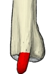 Right ulna (lower end, posterior)
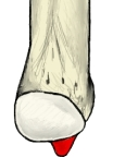 Right ulna (lower end, anterior)
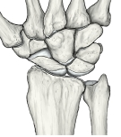 Right wrist (posterior)
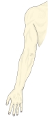 Right upper limb, surface (posterior)
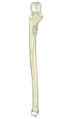 Right ulna (anterior)
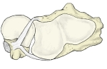 Right radius and ulna (pronated, inferior view)
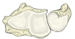 Left radius and ulna (supinated, inferior view)
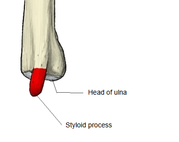
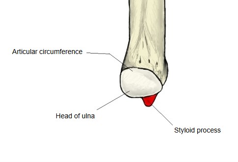
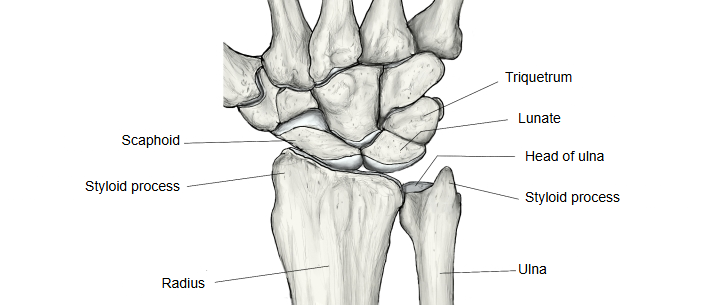
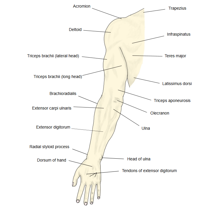
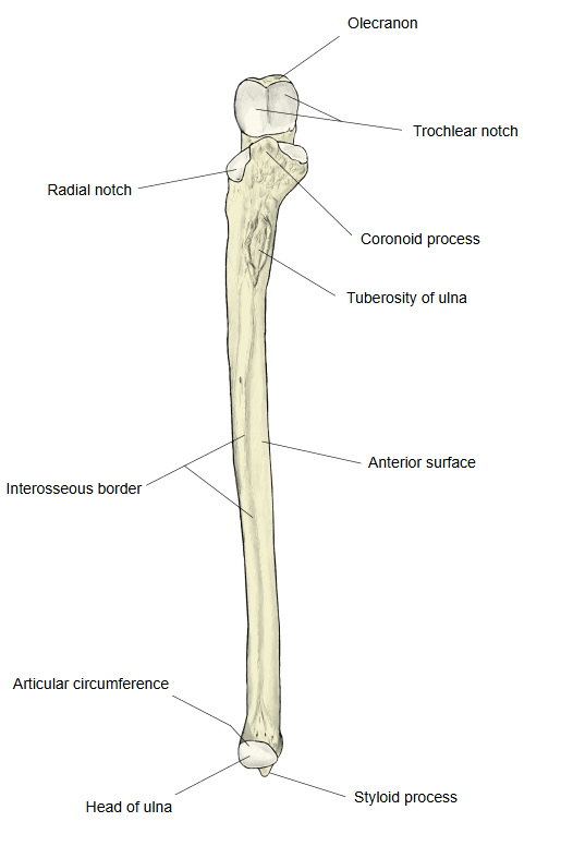
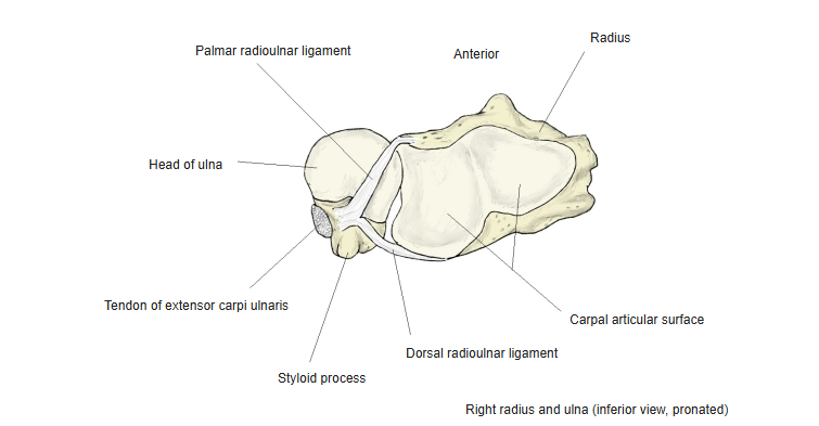
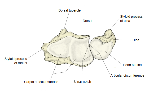
It reaches a little lower than the head, but ends about 1 cm higher than the styloid process of the radius. Its rounded tip gives attachment to the ulnar collateral ligament of the wrist. The head and the styloid process are separated below by a small depression, where the apex of the triangular articular disc of the distal radioulnar joint is attached, and behind by a shallow groove for the tendon of extensor carpi ulnaris. A fracture of the ulnar styloid often accompanies a fracture of the lower end of the radius; it usually needs no separate treatment unless the distal radioulnar joint is unstable.
· Video on the ulna (the styloid process is shown and explained):
Reference websites
[Wikipedia]
· Ulnar styloid process, Wikipedia.
· Ulna, Wikipedia.
[Websites with illustrations]
· Bones of the left forearm, anterior aspect (Gray's Anatomy, fig. 213) (Wikimedia Commons)

· Bones of the left forearm, posterior surface (Gray's Anatomy, fig. 214) (Wikimedia Commons)

· The ulna: proximal, shaft and distal (TeachMeAnatomy)
Where it is found
Related terms
- Head of ulna
- Distal epiphysis of ulna
- Articular circumference of ulna
- Styloid process of radius
- Distal radioulnar joint
- Radiocarpal joint
References and sources
- Further reading: Ulnar styloid process, Wikipedia.
- Further reading: Ulna, Wikipedia.
- Federative International Programme for Anatomical Terminology (FIPAT). Terminologia Anatomica, 2nd ed. (TA2), 2019.