Articular disc (Latin: discus articularis)
A plate of fibrocartilage that completely divides a joint into two; it is found in the temporomandibular joint, the sternoclavicular joint and some other joints. Its edge is attached all round to the joint capsule, so the joint has two separate cavities.
More details
An articular disc is a complete plate that divides the joint in two; when the plate is not complete but ring- or crescent-shaped, it is called an articular meniscus, as in the knee joint. The central part of an articular disc has no blood vessels or nerves; its outer edge receives them from the capsule, and sources differ on how far they reach into it.
that divides
the joint cavity
- Articular disc : a complete plate that divides the joint completely; e.g. the temporomandibular joint
- Articular meniscus : = semilunar cartilage; crescent-shaped, divides the joint incompletely; e.g. the knee joint: medial meniscus and lateral meniscus
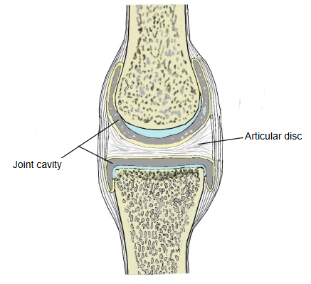
Shape
Thin in the centre and thicker at the edge.
Structure
· Superficial layer: like fibrocartilage, with many oval cells that look like chondrocytes.
· Deep layer: like tendon, with cells that resemble tenocytes.
Function
· Makes the movement of the joint smooth: it improves the fit between the joint surfaces and lets the two compartments move in different ways.
· Cushions the shocks on the joint and spreads the load.
Where it is found
Examples: the temporomandibular joint, the sternoclavicular joint and the distal radioulnar joint; the acromioclavicular joint often has an incomplete one.
Reference websites
[Wikipedia]
· Articular disk, Wikipedia.
[Websites with illustrations]
· Simple, complex (with an articular disc) and compound synovial joints (from Gray's Anatomy) (criticalcaremcqs.com)

· Temporomandibular joint with its articular disk (ahcnweb.net)

Related terms
- Temporomandibular joint
- Sternoclavicular joint
- Distal radioulnar joint
- Acromioclavicular joint
- Lateral meniscus
- Knee joint
- Synovial membrane
References and sources
- Further reading: Articular disk, Wikipedia.
- Federative International Programme for Anatomical Terminology (FIPAT). Terminologia Anatomica, 2nd ed. (TA2), 2019.