Latin: nervi intercostales
Overview
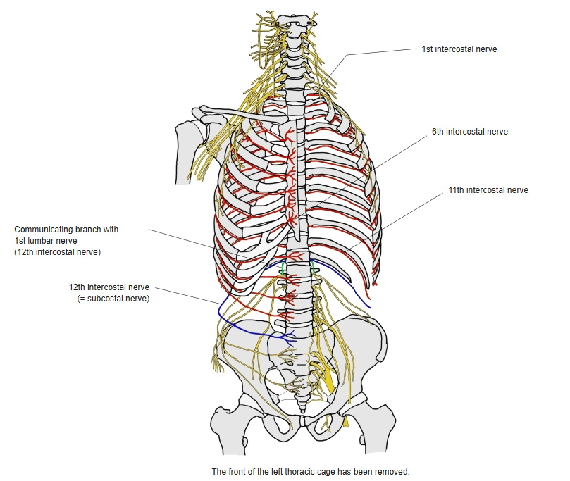
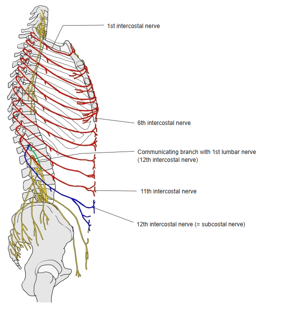
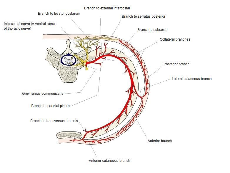
- The ventral rami of T1–T11; motor and sensory, to the chest wall and the front of the abdominal wall.
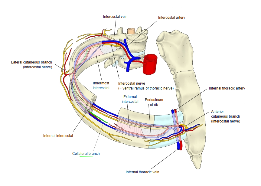
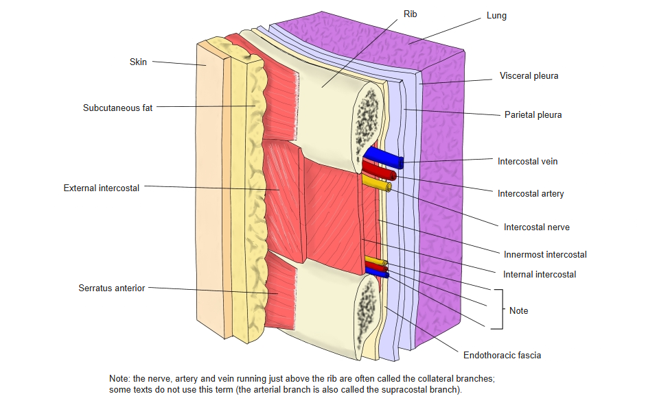
- Each runs along the lower border of the rib above, with the intercostal artery and vein, between the external and internal intercostal muscles.
- The 1st–6th run forwards to the edge of the sternum; the 7th–11th slope down and forwards between internal oblique and transversus abdominis to the linea alba.
- The ventral ramus of T12 runs below the last rib and is the subcostal nerve (sometimes called the 12th intercostal nerve).
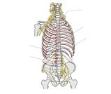
Intercostal nerves (anterior view) 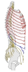
Intercostal nerves (lateral view)
Branches
| Branch | Destination |
|---|---|
| Communicating branches | Sympathetic trunk |
| Collateral branch | Along the upper border of the rib below; intercostal muscles |
| Muscular branches | Muscles of the chest wall and the front and side of the abdominal wall |
| Lateral cutaneous branches | Skin of the side of the chest and abdomen |
| Anterior cutaneous branches | Skin of the front of the chest and abdomen |
| Pleural branches | Parietal pleura; the lower nerves also reach the parietal peritoneum |
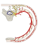
Branches of an intercostal nerve 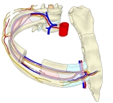
Intercostal nerve and collateral branch 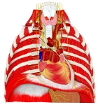
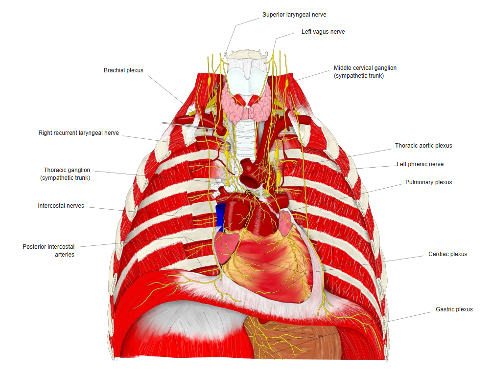
Nerves of the thorax (anterior view) 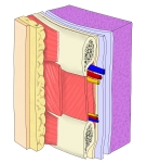
Layers of the thoracic wall 
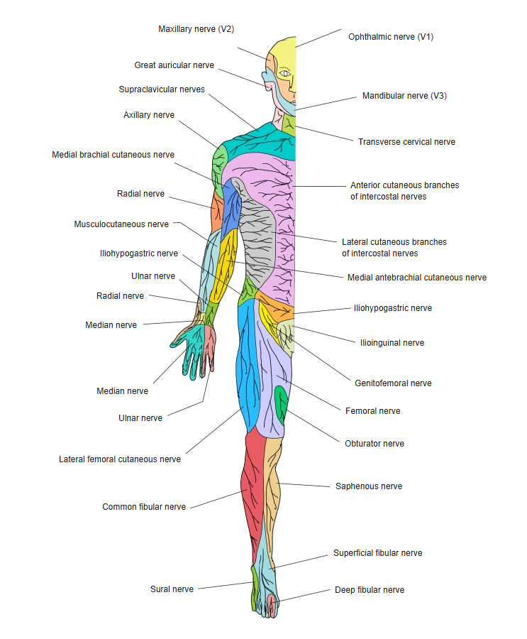
Cutaneous nerves of the body (front) 
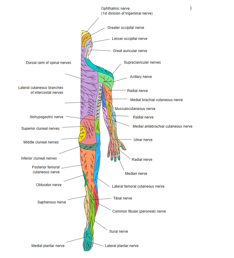
Cutaneous nerves of the whole body (back)
References and sources
- Further reading: Intercostal nerves, Wikipedia.