Latin: rami alveolares superiores anteriores
Overview
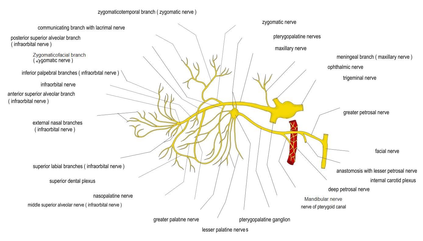
- Branches of the infraorbital nerve (maxillary nerve).
- Supply the upper incisor and canine teeth, their gums, and the mucosa of the front of the nasal cavity.
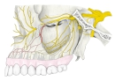
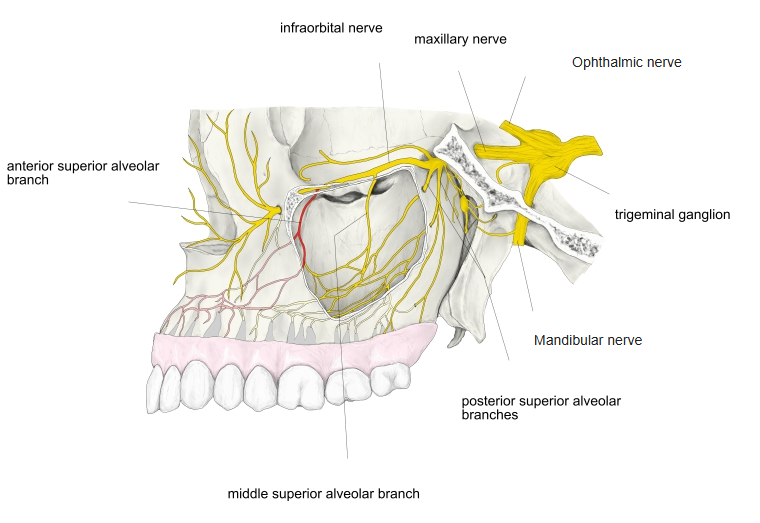
Superior alveolar nerves 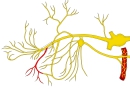
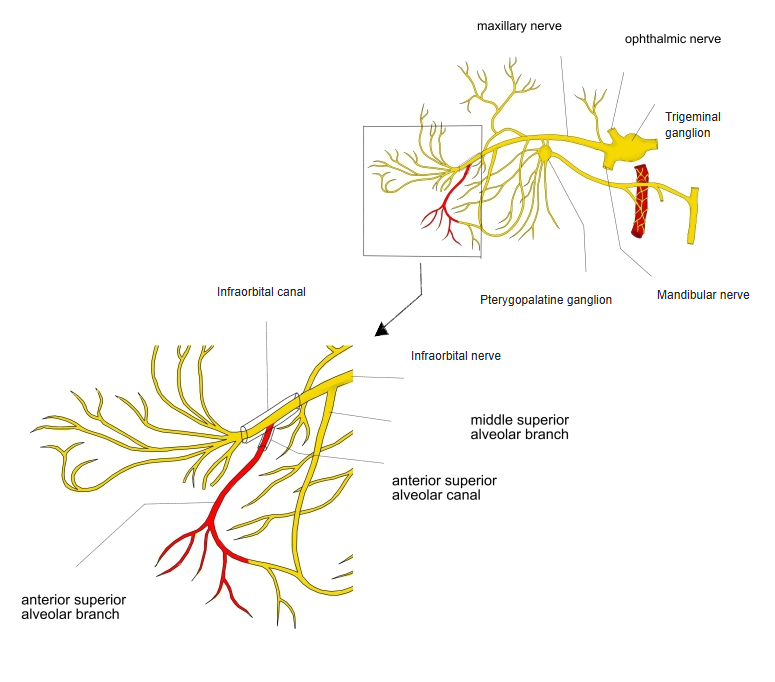
Anterior superior alveolar branch
Course and distribution
- Leave the infraorbital nerve about the middle of the infraorbital canal.
- Descend in a small canal in the front wall of the maxillary sinus (canalis sinuosus).
- A nasal branch reaches the floor and lateral wall of the front of the nasal cavity and the nasal septum.
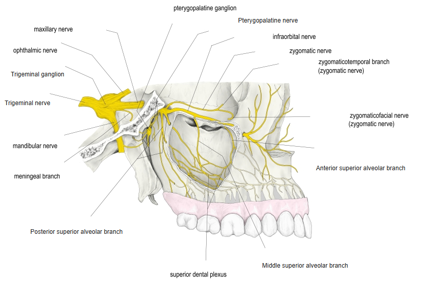
- The dental branches join the middle superior alveolar nerve to form the superior dental plexus, which gives branches to the teeth, gums, alveoli and mucosa.
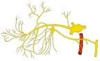
Branches of the maxillary nerve 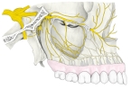
Maxillary nerve, lateral view 
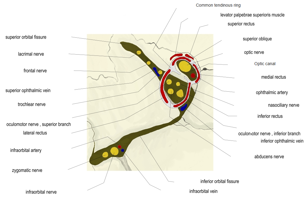
Orbital fissures and optic canal
References and sources
- Further reading: Anterior superior alveolar nerve, Wikipedia.