Sagittal and coronal sections of the nose, mouth, tongue and pharynx. Select a figure to open a larger version with labels.
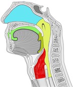
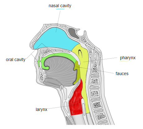
Midsagittal section of the nasal cavity, oral cavity, pharynx and larynx - Colour-coded spaces: nasal cavity (blue), oral cavity (green), fauces (olive), pharynx (yellow) and larynx (red).
- The soft palate separates the nasopharynx from the oral cavity.
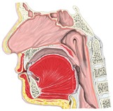
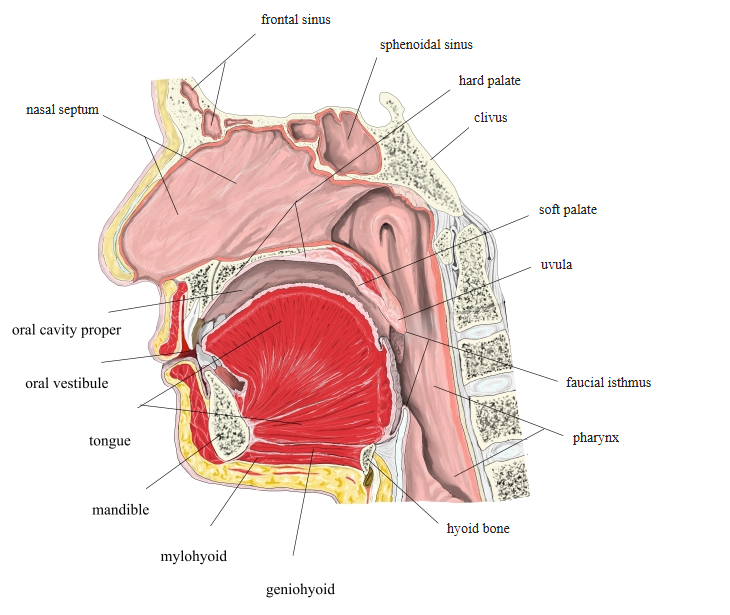
Midsagittal section of the nasal cavity, oral cavity and pharynx - The muscle fibres of the tongue fan out from the genioglossus towards its upper surface.
- The hard palate and soft palate form the roof of the mouth; the cervical vertebrae lie behind the pharynx.
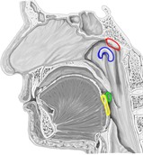
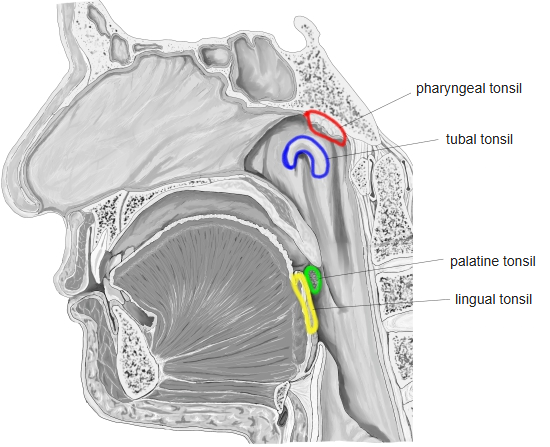
Midsagittal section showing the tonsils - Coloured outlines mark the tonsils of the pharyngeal lymphoid ring: pharyngeal, tubal, palatine and lingual tonsils.
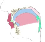
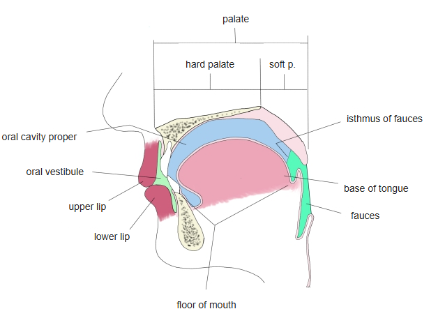
Oral cavity (schematic sagittal section) - Simplified diagram of the lips, palate, tongue and the space of the oral cavity.
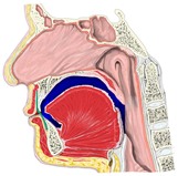
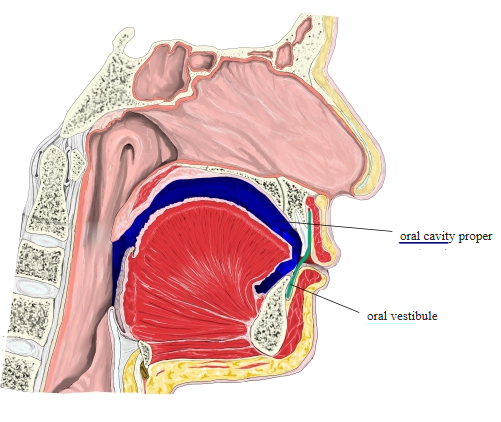
Oral vestibule and oral cavity proper (midsagittal section) - Green: the oral vestibule, between the lips and the teeth.
- Blue: the oral cavity proper, inside the dental arches and above the tongue.
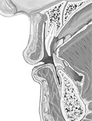
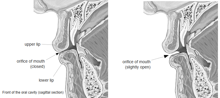
Lips and front of the oral cavity (sagittal section) - Upper and lower lips with the orbicularis oris muscle.
- Incisor teeth, the opening of the mouth and the oral vestibule.
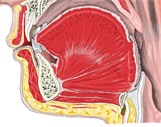
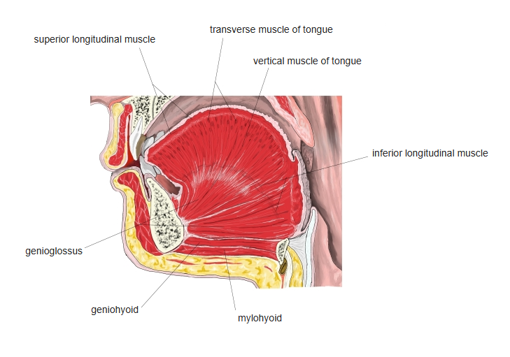
Sagittal section of the oral cavity and tongue - Intrinsic and extrinsic muscles of the tongue; the genioglossus arises from the mandible.
- Geniohyoid and mylohyoid in the floor of the mouth, above the hyoid bone.
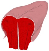
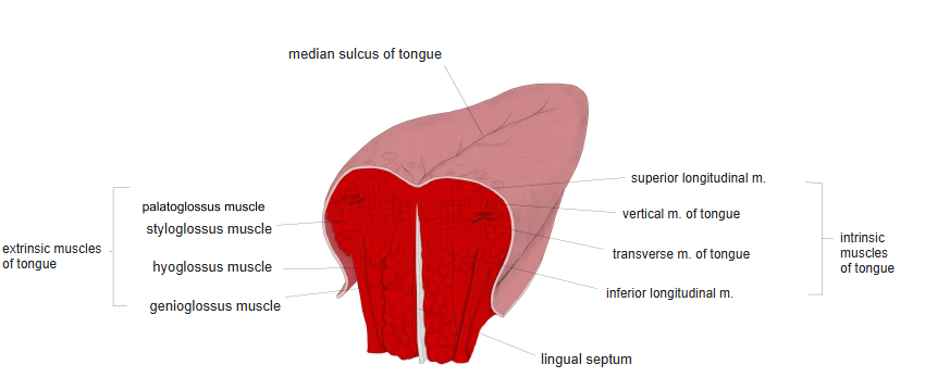
Coronal section of the tongue - A median septum (white) divides the tongue into right and left halves.
- Intrinsic muscles run lengthwise (superior longitudinal), across (transverse) and up and down (vertical).
Back to Cross-sections (all regions)
Other regions: Neck and larynx · Thorax and abdomen · Pelvis and perineum · Upper limb · Lower limb