Sections of the trunk: the diaphragm, abdominal cavity and back, the gallbladder and a lung alveolus. Select a figure to open a larger version with labels.
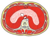
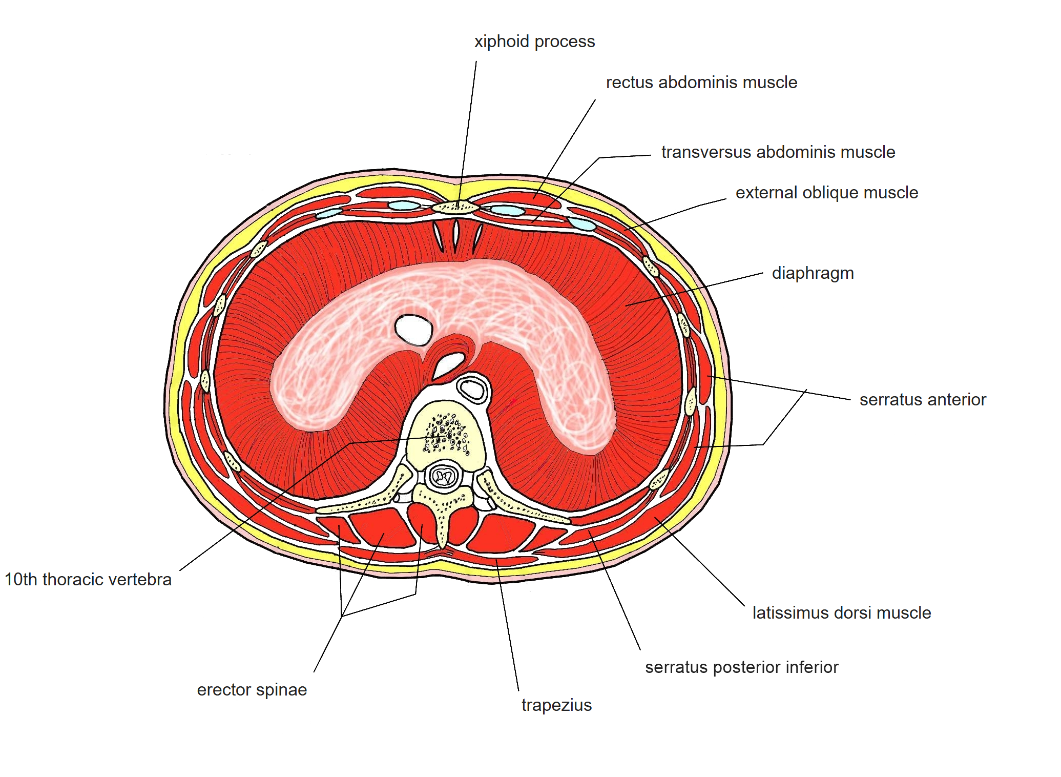
Transverse section at T10: the diaphragm 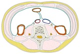
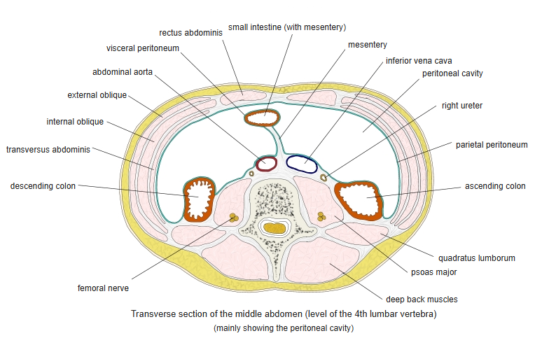
Transverse section of the mid-abdomen - The parietal peritoneum (green line) lines the inside of the abdominal wall.
- Ascending and descending colon lie behind the peritoneum, beside psoas major and quadratus lumborum.
- Aorta (red) and inferior vena cava (blue) in front of the lumbar vertebra.
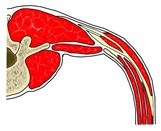
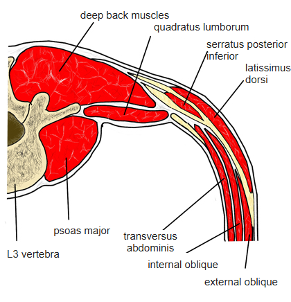
Transverse section of the back at L3 (one side) - Deep back muscles (iliocostalis lumborum and other parts of the erector spinae) behind the transverse process.
- Psoas major beside the vertebral body and quadratus lumborum lateral to it.
- Flat muscles of the abdominal wall: external oblique, internal oblique and transversus abdominis.
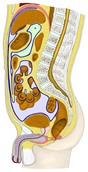
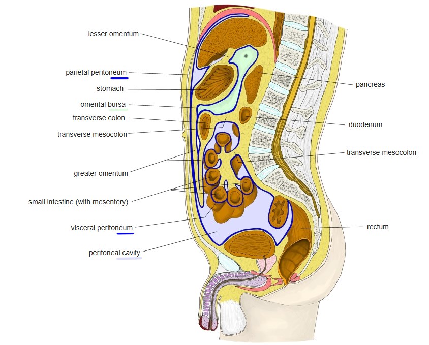
Sagittal section of the abdomen and pelvis: the peritoneal cavity - Blue line: the peritoneum; lavender: the peritoneal cavity; pale green: the omental bursa.
- Liver, stomach, transverse colon, small intestine, urinary bladder and rectum.
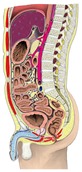
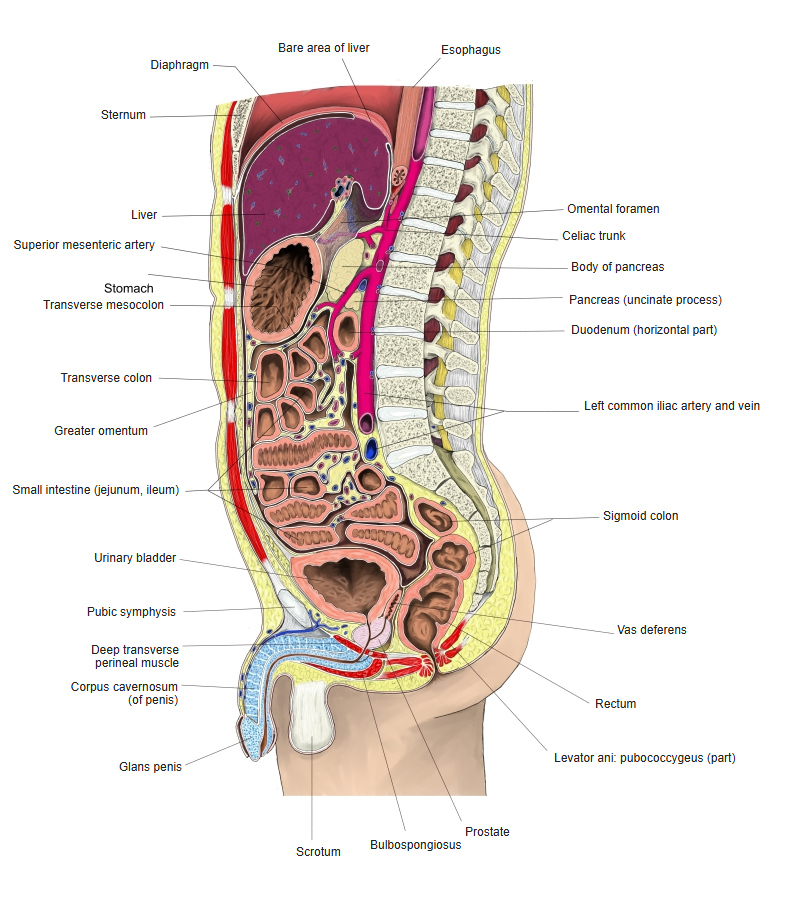
Sagittal section of the abdomen and pelvis (male) - Liver, stomach, pancreas, small intestine and colon in front of the vertebral column.
- Abdominal aorta with its anterior branches.
- Urinary bladder, prostate, rectum and penis; rectus abdominis in the front wall.
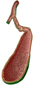
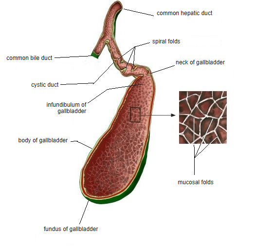
Section of the gallbladder and cystic duct - Fundus, body and neck of the gallbladder with its honeycomb-like mucosa.
- Spiral folds in the cystic duct, which joins the common hepatic duct.
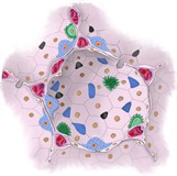
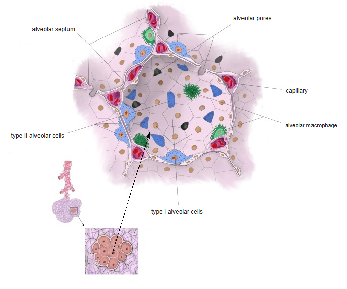
Section of a lung alveolus - Alveolar wall with capillaries containing red blood cells.
- Alveolar epithelial cells and alveolar macrophages on the inner surface.
Back to Cross-sections (all regions)
Other regions: Oral cavity, tongue and pharynx · Neck and larynx · Pelvis and perineum · Upper limb · Lower limb