Latin: musculus scalenus anterior
Overview
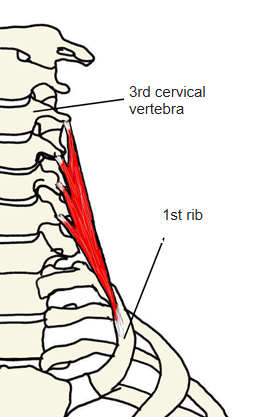
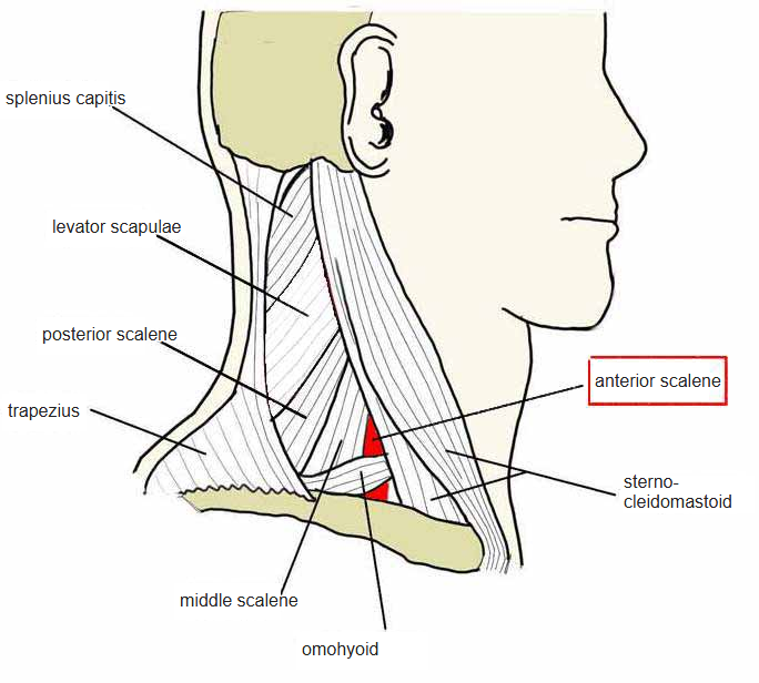
- The most anterior of the scalene muscles, deep in the side of the neck.
- Lies under the sternocleidomastoid; its lower part is crossed by the inferior belly of the omohyoid.
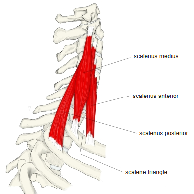
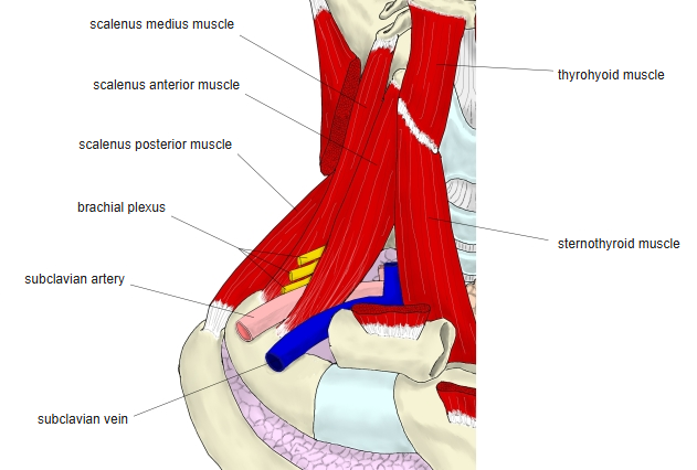
- Near the insertion it leaves a triangular gap with the middle scalene, the scalene hiatus, through which the subclavian artery and brachial plexus pass.
- Continuous with the middle scalene.
Position in the classification
Neck › Deep muscles of the neck › Scalene muscles
- Scalenus anterior
- Scalenus medius
- Scalenus posterior
- Scalenus minimus
Illustrations
Origin / insertion
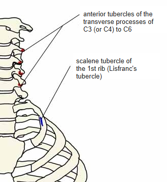
- Origin: anterior tubercles of the transverse processes of C3 (or C4) to C6
- Insertion: scalene tubercle of the 1st rib (Lisfranc's tubercle)
Action
- Raises the 1st rib
- With the ribs fixed, bends the neck forward (and to the same side)
Innervation
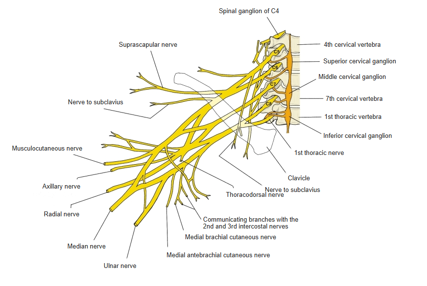
- Branches of the cervical and brachial plexuses (C4–C6/7); English sources: anterior rami of C5–C6
Blood supply
- Not stated in the sources used here.
Related terms
- Scalene muscles
- Middle scalene
- Posterior scalene
- Sternocleidomastoid
- Omohyoid
- First rib
- Subclavian artery
- Brachial plexus
References and sources
- Further reading: Anterior scalene muscle, Wikipedia.