Latin: musculus scalenus medius
Overview
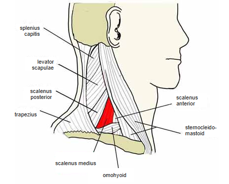
- The largest and longest of the scalene muscles (Wikipedia).
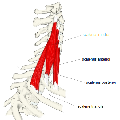
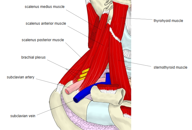
- Near its insertion it leaves a triangular gap with the scalenus anterior, the scalene gap, through which the subclavian artery and the brachial plexus pass.
- Continuous in places with the scalenus anterior and posterior.
Position in the classification
Neck › Deep muscles of the neck › Scalene muscles
- Scalenus anterior
- Scalenus medius
- Scalenus posterior
- Scalenus minimus
Illustrations
Origin / insertion
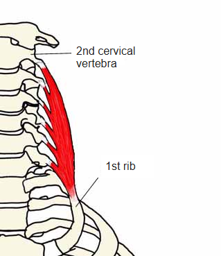
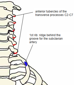
- Origin: anterior tubercles of the transverse processes of C2–C7
- One source gives all cervical vertebrae
- English sources often give the posterior tubercles
- Insertion: ridge on the first rib behind the groove for the subclavian artery
Action
- Raises the first rib
- With the ribs fixed, bends the neck forwards
Innervation
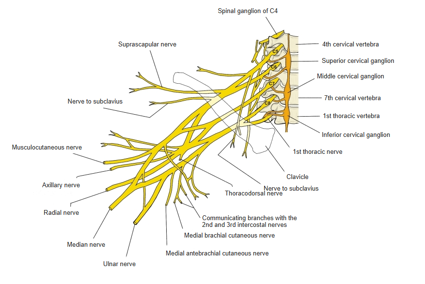
- Direct branches of the cervical plexus and brachial plexus (C2/C3–C8)
Blood supply
- Not given in the sources used here
Related terms
- Anterior scalene
- Posterior scalene
- First rib
- Brachial plexus
- Cervical plexus
- Subclavian artery
References and sources
- Further reading: Scalene muscles, Wikipedia.