Latin: anus
Overview
- The external opening of the digestive tract at the lower end of the anal canal.
- Lies in the anal triangle of the perineum, about 4 cm in front of and below the tip of the coccyx.
- Normally closed by the tonic contraction of the anal sphincters; it opens during defecation.
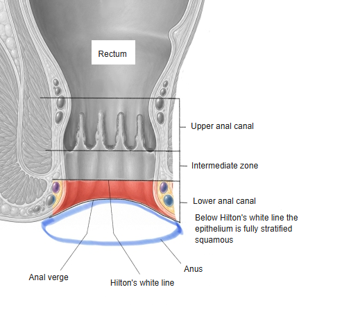
Lower anal canal and anus
Structure
- Anal verge: the margin where the lining of the anal canal meets the perianal skin.
- Skin: keratinised stratified squamous epithelium with hair, sweat glands (including apocrine glands) and sebaceous glands; pigmented and radially folded by the corrugator cutis ani.
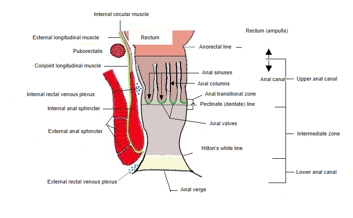
- Internal anal sphincter: thickened circular smooth muscle; involuntary, provides most of the resting tone.
- External anal sphincter: skeletal muscle (subcutaneous, superficial and deep parts); voluntary, blends with the puborectalis above.
- Ischioanal fossae: fat-filled spaces on either side that allow the canal to expand.
Anal canal (coronal section)
Function
- Continence: internal sphincter tone, the external sphincter, the puborectalis sling (anorectal angle) and the venous cushions together keep the anus closed.
- Defecation: rectal filling relaxes the internal sphincter (rectoanal inhibitory reflex); the external sphincter and puborectalis relax voluntarily, and abdominal pressure expels the feces.
Blood supply, lymph and nerves
- Arteries: inferior rectal arteries from the internal pudendal artery.
- Veins: the external rectal venous plexus drains through the inferior rectal veins to the internal pudendal and internal iliac veins.
- Lymph: to the superficial inguinal lymph nodes.
- Nerves: inferior rectal nerves from the pudendal nerve (S2–S4) – sensation of the perianal skin (very sensitive) and motor supply of the external anal sphincter.
References and sources
- Further reading: Anus, Wikipedia.