Rectum (Latin: rectum)
- Overview
- Divisions
- Ampulla of the rectum
- Two flexures
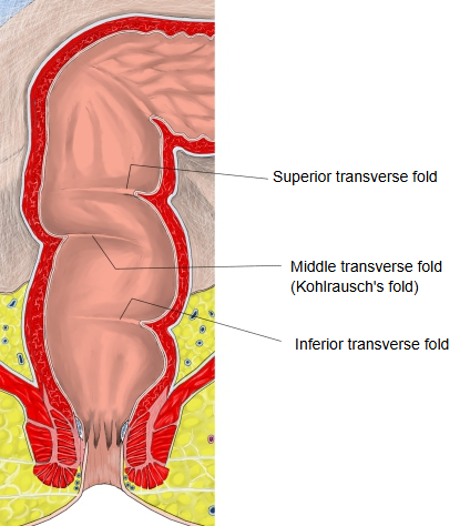
- Three folds
- Relation to the peritoneum (other related membranes)
- Structure of the wall
- Vessels and nerves (arteries, veins, lymphatics, nerves)
- Related terms
- References
Overview
· Some anatomy textbooks count the anal canal as part of the rectum; this site describes the two separately.
· The colon passes gradually into the rectum; the border between them lies roughly at the middle of the sacrum.
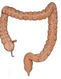
large intestine
- Cecum : length 6–9 cm; the beginning of the large intestine; temporarily stores the contents from the small intestine.
- Vermiform appendix : a tube about 6–10 cm long; helps to keep the balance of the gut bacteria and takes part in immune function.
- Colon : the main part of the large intestine; functions: absorption of water and electrolytes, storage and transport of faeces. Parts: ascending colon (20–25 cm), transverse colon (40–60 cm), descending colon (20–30 cm), sigmoid colon (30–40 cm).
- Rectum : length 12–15 cm; stores faeces temporarily and passes them to the anal canal for defecation.
- Anal canal : length 3–4 cm; the last part of the digestive tract; faeces are expelled through its end, the anus.
[Changes in the faeces]
| Part | State of the faeces | Water | Main features | |
|---|---|---|---|---|
| 1 | Cecum to ascending colon | liquid to porridge-like | very much | contents of the small intestine flow in; fermentation begins |
| 2 | Transverse colon | mushy (semi-fluid) | much | absorption of water proceeds; active gas production |
| 3 | Descending colon | soft solid | moderate | the faeces take shape |
| 4 | Sigmoid colon | solid | little | strong storing function |
| 5 | Rectum | solid (finished faeces) | little | final storage before defecation |
Divisions
| Division | Part | Range and peritoneum | |
|---|---|---|---|
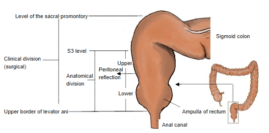
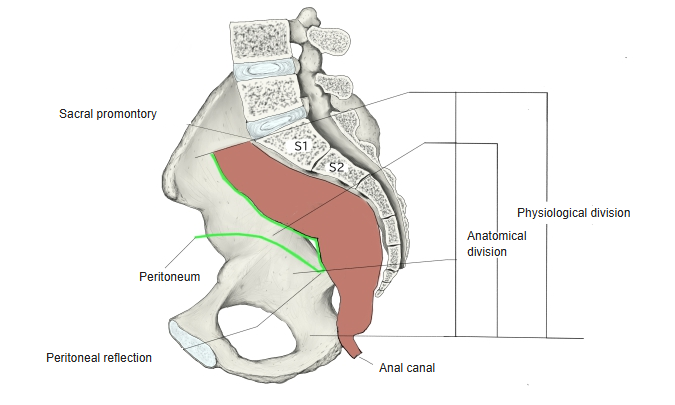
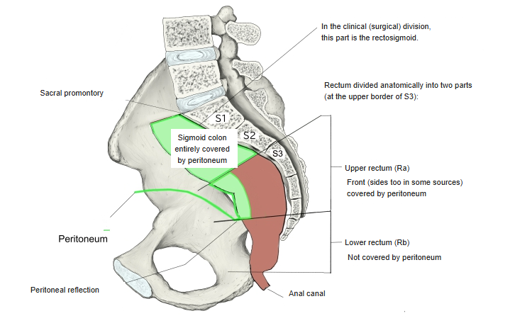
| Clinical | 1 | Rectosigmoid | Range: promontory (sacrum) to the upper border of the 3rd sacral vertebra. Peritoneum: covered all round ⇒ intraperitoneal organ |
| Anatomical | 2 | Upper rectum | Range: upper border of the 3rd sacral vertebra to the peritoneal reflection (about the front of the 4th sacral vertebra). Peritoneum: the front (in some sources also the sides) is covered ⇒ (secondarily) retroperitoneal organ |
| 3 | Lower rectum | Range: peritoneal reflection to the upper end of the levator ani. Peritoneum: not covered at all ⇒ extraperitoneal organ |
[Ampulla of the rectum]
It takes up roughly the lower two-thirds of the rectum and is about 5–8 cm long.
[Two flexures]
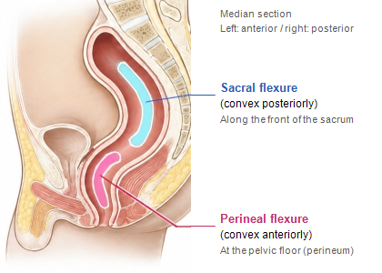
■ Sacral flexure ■
· Position: the upper part of the rectum.
· Curve: bulges backwards.
· Feature: follows the hollow of the sacrum and coccyx at the back of the pelvis, curving backwards close against them.
■ Perineal flexure ■
· Position: the lower part of the rectum, just above the anal canal.
· Curve: bulges forwards (towards the abdomen).
· Feature: formed near the tip of the coccyx, where the puborectalis pulls the rectum forwards.
[Three folds]
folds of
the rectum
- Superior transverse fold : the uppermost fold, on the left wall.
- Middle transverse fold : the best-developed fold, on the right wall; it lies roughly at the level of the peritoneal reflection. = Kohlrausch's fold
- Inferior transverse fold : the lowest fold, on the left wall.
Relation to the peritoneum
Unlike other organs, the relation between the rectum and the peritoneum is a little special.
peritoneum
- Rectosigmoid : covered all round by peritoneum. ⇒ intraperitoneal organ
- Upper rectum : the front (+ sides?) is covered by peritoneum. ⇒ retroperitoneal organ
- Lower rectum : not covered by peritoneum. ⇒ extraperitoneal organ
[Other related membranes]
| Position | Name | Description | |
|---|---|---|---|
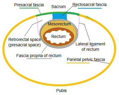
| Around | 1 | Fascia propria of the rectum | (part of the visceral pelvic fascia) wraps the whole mesorectum from outside. |
| 2 | Mesorectum | Called a "meso", but in fact it is the fat around the rectum, containing vessels and lymphatic tissue; unlike the other, fibrous membranes. | |
| 3 | Peritoneum | Covers all or part of the circumference of the upper rectum. | |
| Behind | 1 | Presacral fascia | Covers the front of the sacrum. |
| 2 | Rectosacral fascia | Partly connects the presacral fascia and the fascia propria of the rectum. | |
| 3 | Parietal pelvic fascia | A fascia lining the whole inside of the pelvis; behind, it is continuous with the presacral fascia. | |
| In front | 1 | Denonvilliers' fascia | (male) between the rectum and the prostate and seminal vesicles. |
| 2 | Rectovaginal septum | (female) between the rectum and the vagina. | |
| Below | 1 | Fascia of the levator ani | Covers the surface of the levator ani. |
| 2 | Fasciae around the pelvic diaphragm | The group of fasciae of the pelvic floor. |
Structure of the wall
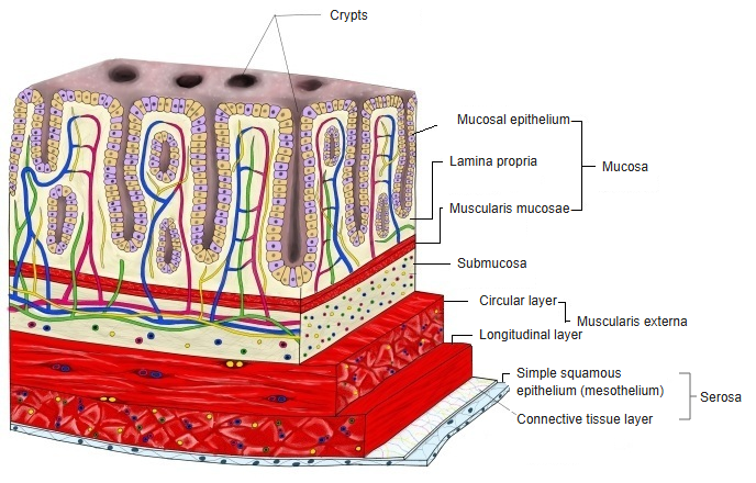
For details, see "Structure of the wall" on the large intestine page.
Vessels and nerves
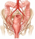
[Arteries]
· Superior rectal artery: the end branch of the inferior mesenteric artery (IMA); the most important artery, supplying most of the rectum (mainly its upper and middle parts).
· Middle rectal artery: arises from the internal iliac artery and supplies the middle and lower parts of the rectum.
· Inferior rectal artery: a branch of the internal pudendal artery; supplies mainly the lower rectum and the anal canal.
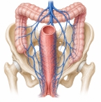
[Veins]
· Superior rectal vein: drains into the portal system (the route to the liver, portal vein).
· Middle and inferior rectal veins: drain into the systemic circulation (the route to the inferior vena cava, through the internal iliac vein).
* This meeting point of the portal and systemic veins is important: in portal hypertension the veins here can become dilated (varices).
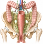
[Lymphatics]
The lymphatics are medically very important as routes of spread of rectal cancer. Lymph flows mainly along the blood vessels.
· Upper rectum: along the superior rectal vessels towards the inferior mesenteric lymph nodes (deep in the abdomen).
· Middle and lower rectum: not only upwards but also sideways, to the internal iliac lymph nodes and others.
· Part near the anus: further down, a route to the inguinal lymph nodes (at the groin) is added.
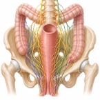
[Nerves]
The rectum is controlled by autonomic nerves, which cannot be moved at will.
· Parasympathetic (pelvic splanchnic nerves): arise from the S2–S4 spinal nerves and promote contraction of the rectum and defecation. The feeling that faeces have collected (the urge to defecate) is also carried by these nerves.
· Sympathetic (hypogastric nerves and others): arise from the lumbar spinal cord (L1–L2) and reach the rectum through the superior hypogastric plexus; they inhibit the movements of the rectum and tighten the internal anal sphincter to hold the faeces.
· Pelvic plexus (inferior hypogastric plexus): a network where these autonomic nerves gather; it runs right beside the rectum. Injury to it during surgery can affect urination and sexual function.
Related terms
Main anatomical terms related to the rectum, in alphabetical order.
| A | Ampulla of the rectum |
| H | Houston's valves (= transverse folds of the rectum) |
| I | Ischioanal fossa |
| K | Kohlrausch's fold (= middle transverse fold of the rectum) |
| M | Middle transverse fold of rectum |
| P | Perineal flexure |
| R | Rectovesical pouch |
| S | Sacral flexure |
| T | Transverse folds of rectum |
References and sources
- Further reading: Rectum, Wikipedia.