Latin: omentum minus
Overview
- Double layer of peritoneum between the liver and the lesser curvature of the stomach and the first part of the duodenum.
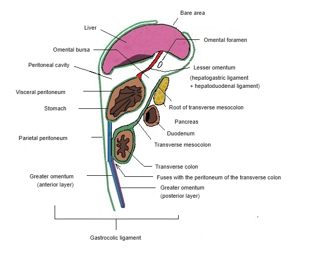
- Very thin; it forms part of the front wall of the omental bursa (lesser sac).
- Its right free border forms the front boundary of the omental (epiploic) foramen.
- The counterpart below the stomach is the greater omentum.
Greater and lesser omentum 
Peritoneal cavity in a median section
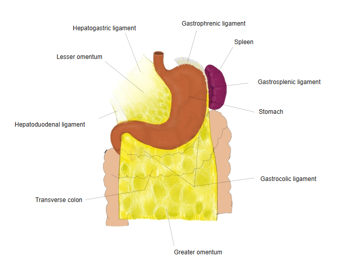
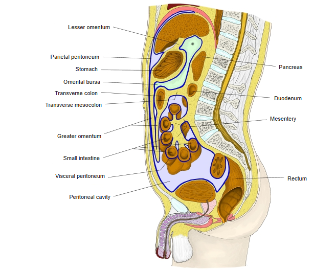
Parts
| Part | Between | Contents |
|---|---|---|
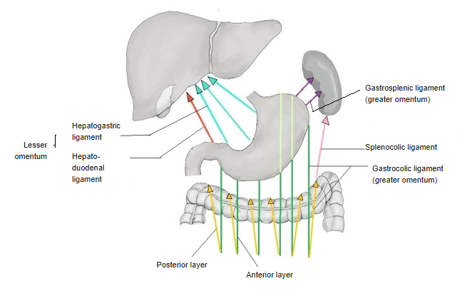
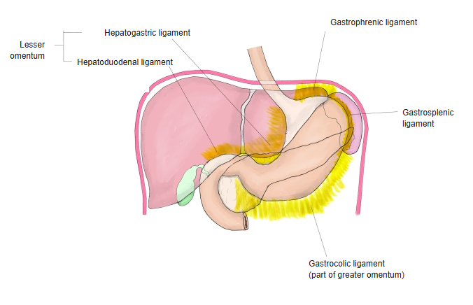
| Hepatogastric ligament | Liver (fissure for the ligamentum venosum) and the lesser curvature | Left and right gastric vessels, gastric lymph nodes, vagal branches |
| Hepatoduodenal ligament | Porta hepatis and the first part of the duodenum (thick right free border) | Proper hepatic artery, common bile duct, hepatic portal vein, lymphatics and the hepatic plexus |
- In the hepatoduodenal ligament the bile duct lies on the right, the hepatic artery on the left and the portal vein behind both.
- Some descriptions add small hepatophrenic and hepatoesophageal parts toward the diaphragm and esophagus.
Parts of the lesser and greater omentum 
Ligaments of the stomach
Relations
- Above, the two layers separate at the porta hepatis; on the left they reach the diaphragm and enclose the end of the esophagus.
- Below, they separate to cover the front and back of the stomach and the first part of the duodenum.
- Behind it lies the omental bursa; the hepatoduodenal ligament can be compressed between finger and thumb through the omental foramen to control bleeding from the liver (Pringle manoeuvre).
Omenta in a sagittal diagram
References and sources
- Further reading: Lesser omentum, Wikipedia.