Latin: peritoneum
Overview
- The serous membrane lining the abdominal and pelvic cavities and covering many of their organs.
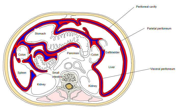
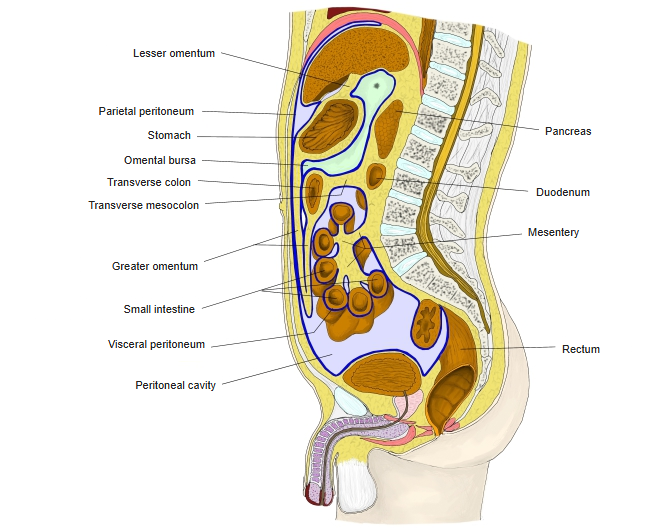
- Parietal peritoneum lines the walls; visceral peritoneum covers the organs; the two are continuous.
- The narrow space between them is the peritoneal cavity, holding only a thin film of serous fluid.
- Double folds of peritoneum – mesenteries, omenta and peritoneal ligaments – connect organs to the wall and to each other and carry their vessels and nerves.
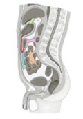
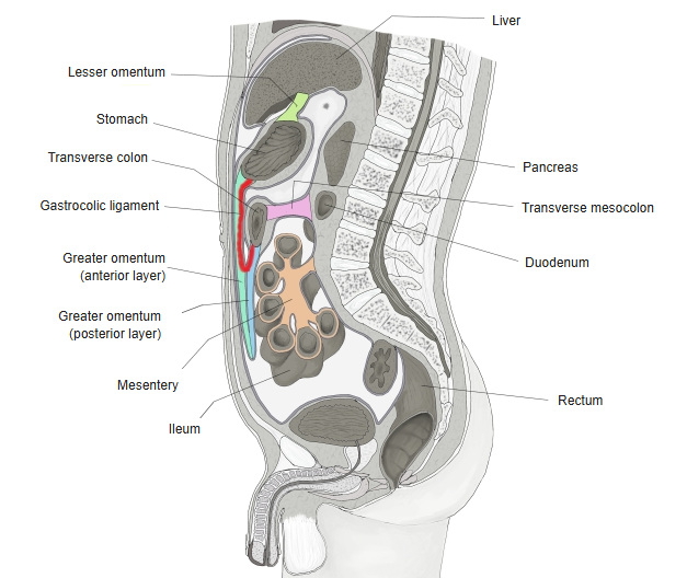
Peritoneum in a median section 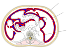
Peritoneum in a transverse section
Peritoneum and the organs
| Group | Description | Examples |
|---|---|---|
| Intraperitoneal organs | Almost entirely covered by peritoneum and suspended by a mesentery or peritoneal fold; usually mobile | Stomach, jejunum, ileum, transverse and sigmoid colon, liver, spleen, first part of the duodenum |
| Primarily retroperitoneal | Develop and stay behind the peritoneum, joined to the posterior wall by connective tissue | Kidneys, suprarenal glands, ureters, abdominal aorta, inferior vena cava, sympathetic trunk |
| Secondarily retroperitoneal | Had a mesentery in the embryo, then fused with the posterior wall; covered only in front | Pancreas (except the tail), duodenum (2nd–4th parts), ascending and descending colon |
Transverse section at the level of L4
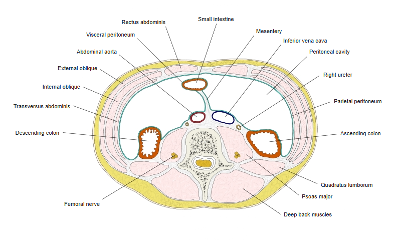
Structure
- Mesothelium: a single layer of flat cells forming the smooth free surface.
- Submesothelial (subserous) layer: loose connective tissue attaching the membrane to the wall or organ.
- Total surface area is roughly the same as the skin (about 1.8 m²).
Folds and spaces
| Structure | Description |
|---|---|
| Mesentery | Double layer attaching an organ to the posterior abdominal wall; see mesentery and the mesocolons |
| Greater omentum | Apron-like fold hanging from the greater curvature of the stomach over the intestines |
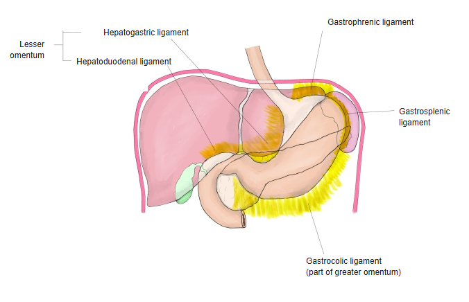
| Lesser omentum | Fold between the liver and the lesser curvature of the stomach and the first part of the duodenum |
| Peritoneal ligaments | Folds between organs or between an organ and the wall, e.g. falciform, gastrosplenic, gastrocolic ligaments |
| Omental bursa (lesser sac) | Space behind the stomach and lesser omentum; opens into the greater sac at the omental (epiploic) foramen |
| Recesses and fossae | Pockets of the peritoneal cavity, e.g. hepatorenal recess, rectouterine pouch (female) or rectovesical pouch (male) |
| Folds of the anterior wall | Median, medial and lateral umbilical folds below the umbilicus |
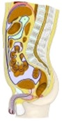
Peritoneal cavity in a median section 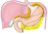
Ligaments of the stomach
Function
- Lubrication: serous fluid lets the organs glide on each other with little friction.
- Support: folds hold the organs in place and carry their blood vessels, lymphatics and nerves.
- Defence: peritoneal fluid and cells remove bacteria and help to wall off infection; the greater omentum tends to move to inflamed areas.
- Fat storage: especially in the greater omentum and mesenteries.
- Absorption and exudation: large surface exchanges fluid rapidly (used in peritoneal dialysis).
Vessels and nerves
- Blood vessels: few in the membrane itself; supplied by vessels of the wall or organ it covers.
- Lymph: superficial and deep lymphatic plexuses.
- Parietal peritoneum: somatic nerves (lower intercostal, subcostal, iliohypogastric, ilioinguinal, phrenic) – pain is sharp and well localised.
- Visceral peritoneum: autonomic (visceral afferent) fibres – sensitive to stretch, not to cutting; pain is dull and poorly localised.
Clinical notes
- Ascites: abnormal fluid in the peritoneal cavity.
- Peritonitis: inflammation of the peritoneum, often from a perforated organ.
References and sources
- Further reading: Peritoneum, Wikipedia.