Latin: uterus; metra
Overview
- Hollow, pear-shaped muscular organ in the middle of the pelvis where the embryo implants and the fetus develops.
- In a non-pregnant adult about 7–8 cm long, 5 cm wide and 2–3 cm thick.
- Lies between the bladder in front and the rectum behind.
- Usually anteverted and anteflexed: tilted forward over the bladder.
Female pelvis (median section)
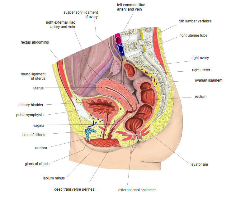
Parts
| Part | Description |
|---|---|
| Fundus | Rounded top above the openings of the uterine tubes |
| Body | Main part; contains the slit-like uterine cavity |
| Isthmus | Short narrow part between body and cervix |
| Cervix | Lower cylindrical part; its lower end projects into the vagina and opens at the external os |
Wall
| Layer | Description |
|---|---|
| Endometrium | Mucosa; its functional layer thickens and is shed each month in the menstrual cycle |
| Myometrium | Thick smooth muscle; contracts in labour |
| Perimetrium | Peritoneal covering |
Support and vessels
- Ligaments: broad ligament (peritoneal fold), round ligament (to the labium majus through the inguinal canal), cardinal (transverse cervical) and uterosacral ligaments; the pelvic floor gives the main support.
- Peritoneal pouches: vesicouterine pouch in front, rectouterine pouch (pouch of Douglas) behind – the lowest point of the peritoneal cavity.
- Artery: uterine artery from the internal iliac artery; it crosses above the ureter near the cervix.
References and sources
- Further reading: Uterus, Wikipedia.