Latin: choroidea
Overview
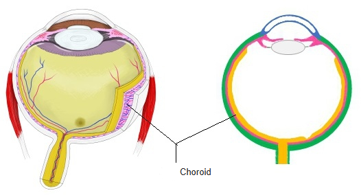
- The posterior part of the vascular layer (uvea), between the retina and the sclera.
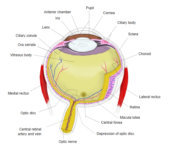
- Extends from the optic nerve forward to the ora serrata, where it continues into the ciliary body.
- Thin: about 0.2 mm at the back of the eye, thinning to about 0.1 mm in front.
- Dark brown from melanin, which absorbs stray light inside the eye.
- Supplies oxygen and nutrients to the outer layers of the retina (photoreceptors and pigment epithelium); around the optic nerve it is firmly fixed to the sclera.
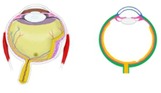
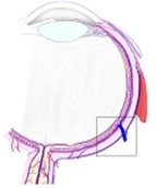
Choroid (diagram) 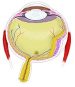
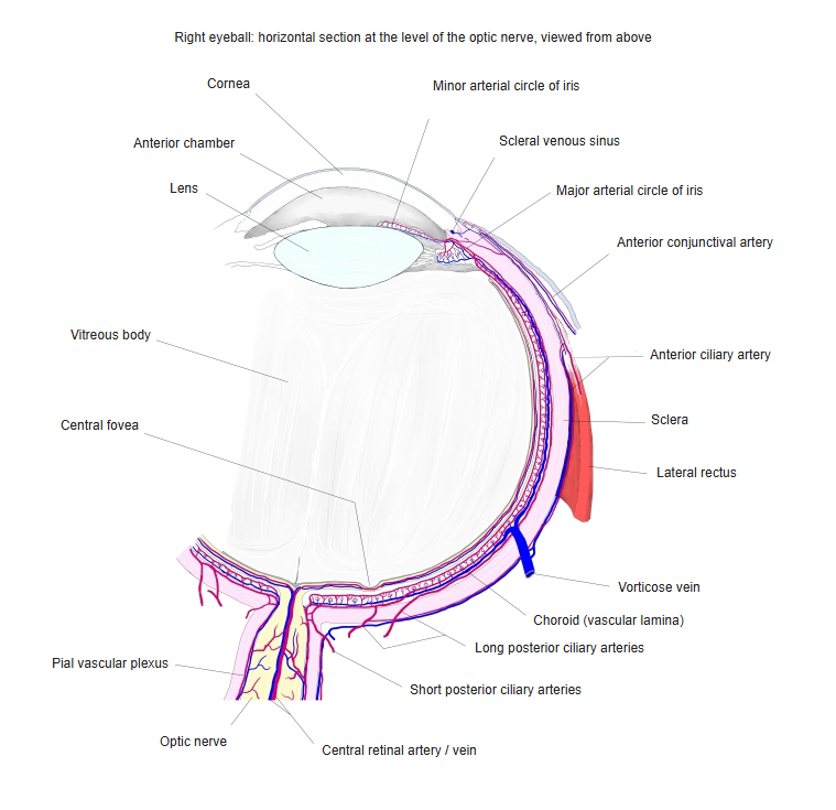
Eyeball, horizontal section
Layers
| Layer (outside to inside) | Features |
|---|---|
| Suprachoroid lamina | Loose pigmented connective tissue next to the sclera; the perichoroidal space lies here |
| Vascular lamina | Large (outer) and medium-sized (inner) arteries and veins in pigmented stroma |
| Choriocapillary lamina | A dense sheet of wide capillaries that feeds the outer retina |
| Basal lamina (Bruch's membrane) | Thin glassy membrane between the capillaries and the retinal pigment epithelium |
Choroid and its vessels
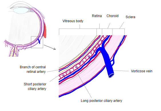
Blood supply
- Arteries: about 15–20 short posterior ciliary arteries pierce the sclera around the optic nerve; branches of the ophthalmic artery.
- Veins: gather into the 4–6 vorticose veins, which leave the eyeball behind the equator and drain into the ophthalmic veins.
- This choroidal (uveal) circulation is separate from the retinal circulation of the central retinal artery.
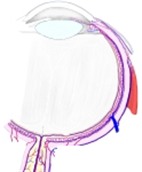
Right eyeball, horizontal section
References and sources
- Further reading: Choroid, Wikipedia.