Latin: sclera
Overview
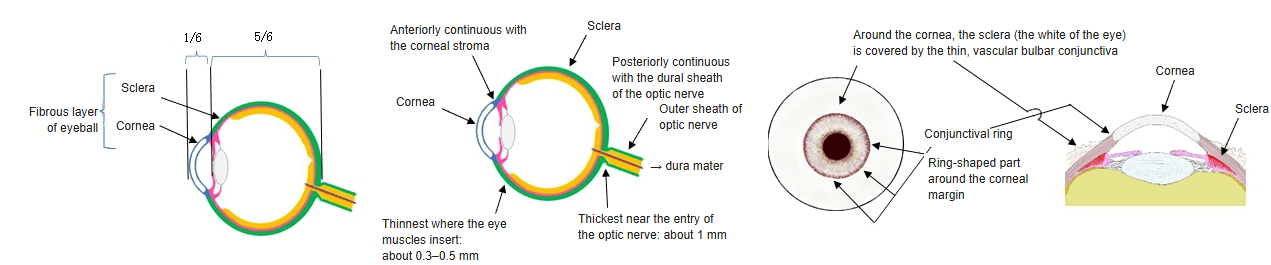
- The white, opaque posterior five-sixths of the fibrous layer – the "white of the eye".
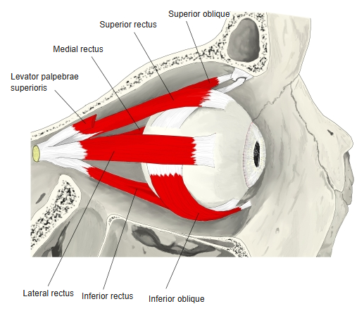
- Dense, irregular collagen with some elastic fibres; it keeps the shape of the eyeball and gives insertion to the extraocular muscles.
- Few blood vessels; bluish in children (thin, the pigment shows through) and slightly yellowish in old age.
- In front it is continuous with the cornea at the corneal limbus; near the limbus the scleral venous sinus (Schlemm's canal) runs around the eye.
- The visible front part is covered by the bulbar conjunctiva.
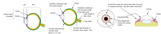
Sclera (diagram) 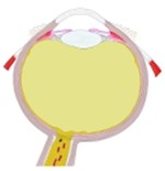
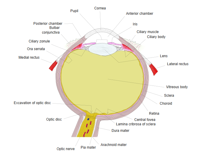
Right eyeball, horizontal section (model)
Thickness and openings
- Thickest at the posterior pole near the optic nerve – about 1 mm.
- About 0.6 mm at the limbus and 0.4–0.5 mm at the equator.
- Thinnest just behind the insertions of the rectus muscles – about 0.3 mm.
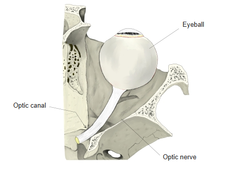
- The optic nerve leaves through the lamina cribrosa, a sieve-like plate; here the outer part of the sclera continues into the dural sheath of the optic nerve.
- Small openings around the optic nerve pass the posterior ciliary arteries and nerves; behind the equator the vorticose veins pass out; near the limbus the anterior ciliary vessels pass in.
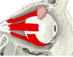
Right extraocular muscles, lateral view 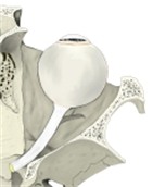
Eyeball and optic nerve in the orbit
Layers
| Layer (outside to inside) | Features |
|---|---|
| Episclera | Loose, vascular connective tissue under the conjunctiva and the fascial sheath of the eyeball |
| Substantia propria (stroma) | Interwoven collagen bundles with a few elastic fibres; the irregular arrangement makes it opaque |
| Lamina fusca | Thin, brownish layer with pigment cells next to the choroid |
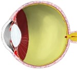
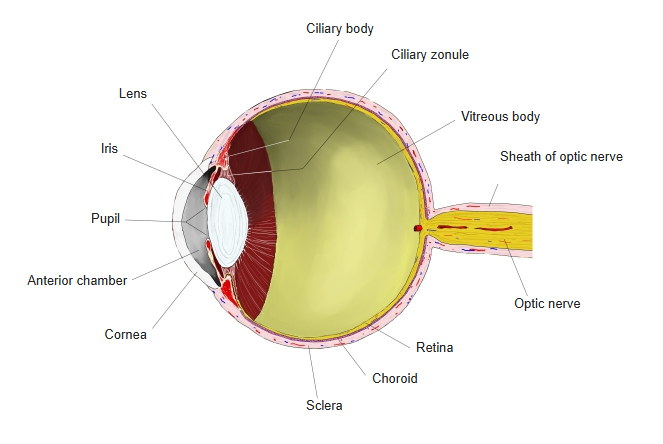
Eyeball, longitudinal section
References and sources
- Further reading: Sclera, Wikipedia.