Latin: cochlea
Overview
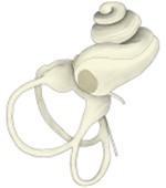
- The snail-shaped front part of the bony labyrinth, containing the organ of hearing.
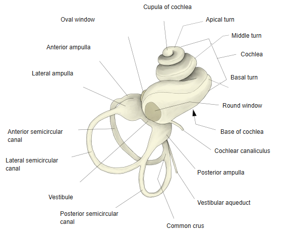
- A spiral canal of about 2.5 turns (basal, middle and apical turns) around a conical bony core, the modiolus; about 30–35 mm long if unrolled.
- Its base faces the internal acoustic meatus; its apex (cupula) points forwards and laterally.
- The basal turn bulges into the tympanic cavity as the promontory.
Right bony labyrinth 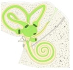
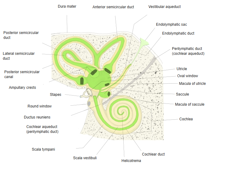
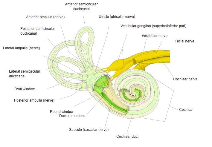
Labyrinth in the temporal bone (schematic)
Internal structure
| Structure | Description |
|---|---|
| Modiolus | Bony core; contains the spiral ganglion and the fibres of the cochlear nerve |
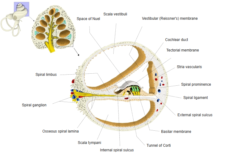
| Osseous spiral lamina | Bony shelf winding around the modiolus; with the basilar membrane it divides the canal |
| Scala vestibuli | Upper perilymphatic channel; begins at the vestibule (oval window) |
| Cochlear duct | Endolymph-filled middle channel containing the spiral organ |
| Scala tympani | Lower perilymphatic channel; ends at the round window |
| Helicotrema | Opening at the apex where the scala vestibuli and scala tympani meet |
- Sound vibrations at the oval window travel through the perilymph of the scala vestibuli and make the basilar membrane vibrate; high tones act near the base, low tones near the apex.
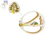
Cochlea, cross-section of one turn
Nerves
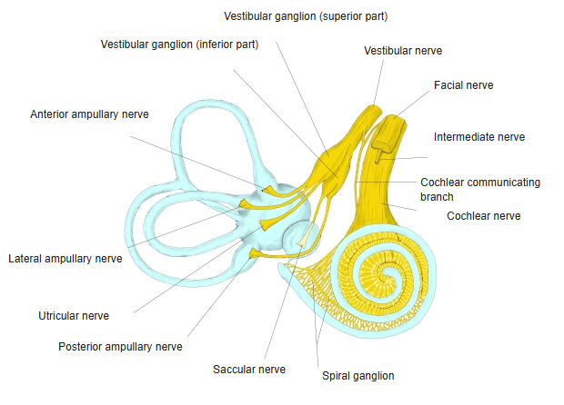
- The bipolar cells of the spiral ganglion in the modiolus send their peripheral processes to the hair cells.
- Their central processes form the cochlear nerve, part of the vestibulocochlear nerve.
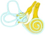
Nerves of the membranous labyrinth 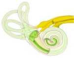
Labyrinth and its nerves
References and sources
- Further reading: Cochlea, Wikipedia.