Latin: discus nervi optici
Overview
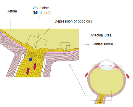
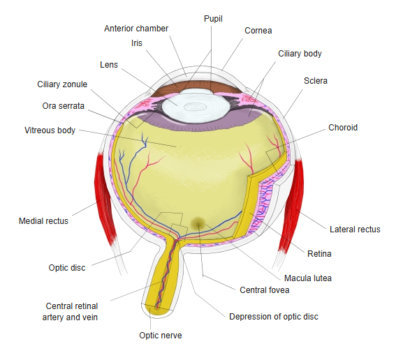
- The spot on the retina where the optic nerve fibres leave the eyeball and the central retinal artery and vein enter.
- Pale, slightly oval area about 1.5–1.8 mm across, about 3–4 mm to the nasal (medial) side of the macula.
- Has no photoreceptors, so it forms the blind spot of the visual field.
- Carries roughly 1–1.2 million ganglion cell axons; they gain their myelin only behind the eyeball.
- Its centre has a small pit, the depression of the optic disc (physiological cup); also called the optic nerve head or optic papilla.
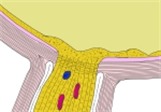
Optic disc and macula, section 
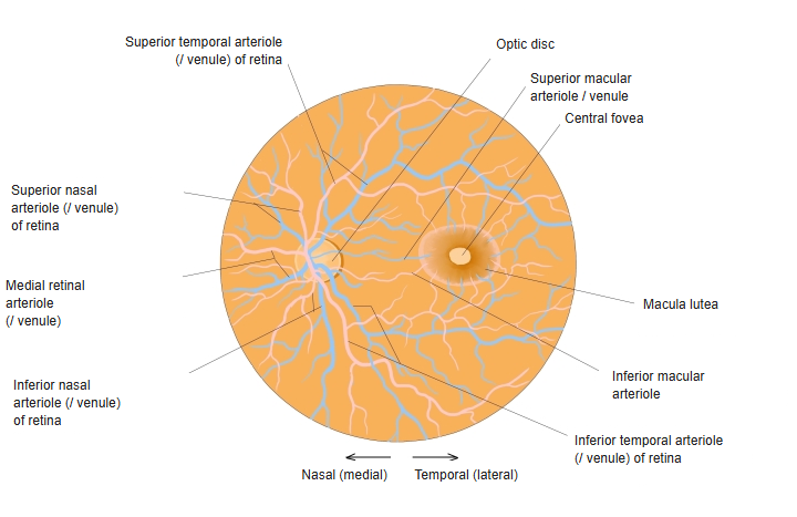
Optic disc and macula in the fundus
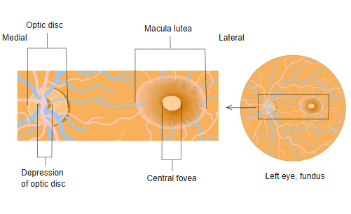
In the fundus
- Seen through an ophthalmoscope as a yellowish-pink disc with a sharp edge.
- The branches of the central retinal artery and vein spread out from it towards the four quadrants of the retina.
- Swelling of the disc (papilloedema) can be a sign of raised intracranial pressure.
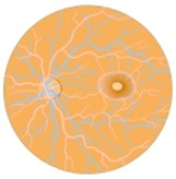
Fundus of the eye 
Eyeball, horizontal section
References and sources
- Further reading: Optic disc, Wikipedia.