Latin: retina
Overview
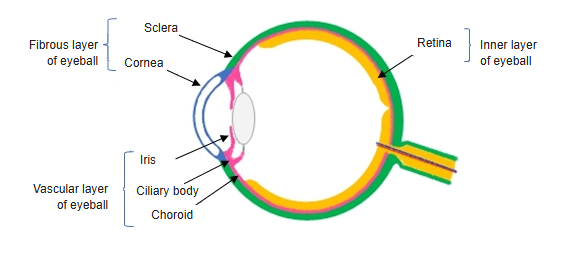
- The inner layer of the eyeball (inner layer): it contains the photoreceptors that turn light into nerve signals.
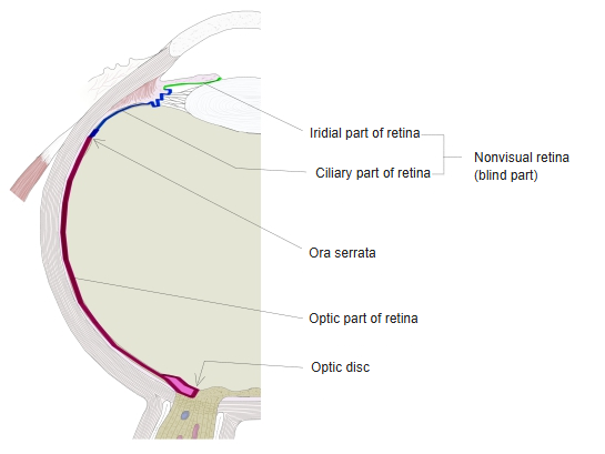
- Two parts: the optic part, which senses light, and the nonvisual (blind) part over the ciliary body and iris; the boundary is the wavy ora serrata.
- Has two layers: the outer pigment epithelium (attached to the choroid) and the inner neural retina; they separate in retinal detachment.
- Its ganglion cell axons converge on the optic disc and form the optic nerve.

Retina and the layers of the eyeball wall 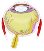
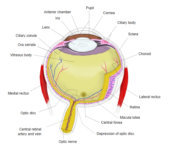
Eyeball, horizontal section
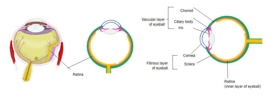
Parts of the retina
| Part | Features |
|---|---|
| Optic part | From the optic disc to the ora serrata; photoreceptors and neurons |
| Ciliary part | Two-layered epithelium on the ciliary body |
| Iridial part | Two-layered pigmented epithelium on the back of the iris |
- The ciliary and iridial parts together form the nonvisual (blind) part.
Parts of the retina 
Layers of the eyeball wall (diagram)
Special regions
| Region | Features |
|---|---|
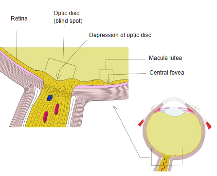
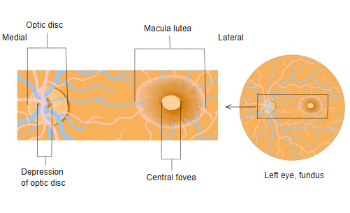
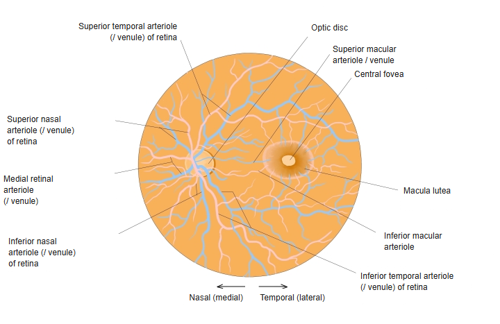
| Optic disc | Where the optic nerve leaves and the central retinal vessels enter; no photoreceptors – the blind spot |
| Macula lutea | Yellowish area lateral (temporal) to the optic disc; its central pit, the central fovea, gives the sharpest vision |
| Ora serrata | The thin, saw-toothed front border of the optic part |
- The central retinal artery supplies the inner layers; its branches spread from the optic disc in four quadrants. The outer layers are fed from the choroid.
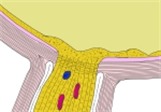
Optic disc and macula, section 
Optic disc and macula in the fundus 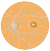
Fundus of the eye
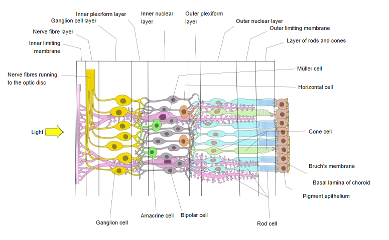
Layers and cells
| Cell | Role |
|---|---|
| Photoreceptors: rods and cones | Turn light into electrical signals; about 120 million rods (dim light) and 6 million cones (colour, detail) |
| Bipolar cells | Pass signals from the photoreceptors to the ganglion cells |
| Horizontal cells | Link photoreceptors sideways and sharpen contrast |
| Amacrine cells | Link bipolar and ganglion cells sideways and shape their signals |
| Ganglion cells | Send the final signals to the brain; their axons form the optic nerve |
| Müller cells | Glial cells that support the retina and span almost its full thickness |
- From the vitreous outwards the classic ten layers are: inner limiting membrane, nerve fibre layer, ganglion cell layer, inner plexiform layer, inner nuclear layer, outer plexiform layer, outer nuclear layer, outer limiting membrane, layer of rods and cones, pigment epithelium.
- Light passes through the whole thickness of the retina before reaching the photoreceptors.
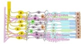
Layers and cells of the retina
References and sources
- Further reading: Retina, Wikipedia.