Latin: Septum interventriculare
Overview
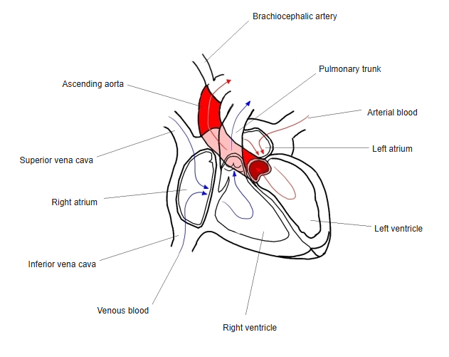
- The wall between the right ventricle and the left ventricle.
- Mostly a thick muscular part; a small thin membranous part lies just below the aortic valve.
Chambers of the heart and the flow of blood
Features
- Bulges into the right ventricle, because pressure is higher on the left.
- Its position on the surface is shown by the anterior and posterior interventricular sulci.
- The atrioventricular bundle runs along the top of the muscular part and divides into the right and left bundle branches.
- Blood supply: septal branches of the anterior and posterior interventricular arteries.
Clinical notes
- Ventricular septal defect (VSD), usually in the membranous part, is the most common congenital heart defect.
References and sources
- Further reading: Interventricular septum, Wikipedia.