Latin: Cor
Overview
- A hollow muscular organ that pumps blood through the body; about the size of a closed fist (roughly 250–350 g in adults).
- Lies in the middle mediastinum, enclosed in the pericardium, with about two-thirds of its mass to the left of the midline.
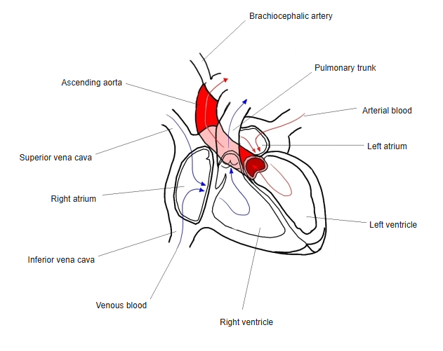
- Has four chambers: the right atrium and right ventricle (pulmonary circulation) and the left atrium and left ventricle (systemic circulation).
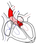
Chambers of the heart and the flow of blood 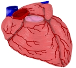
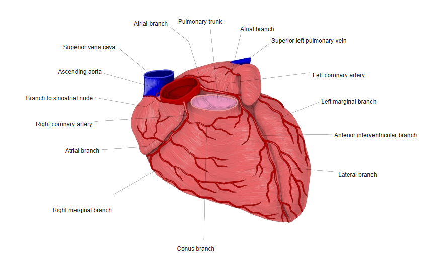
Heart from the front with the coronary arteries
Shape and surfaces
- Apex: formed by the left ventricle; points down, forward and to the left (about the left 5th intercostal space, near the midclavicular line).
- Base: the posterior surface, formed mainly by the left atrium, where the pulmonary veins enter.
- Sternocostal (anterior) surface: mainly the right ventricle. Diaphragmatic (inferior) surface: mainly the left ventricle.
- The coronary sulcus separates the atria from the ventricles; the anterior and posterior interventricular sulci mark the interventricular septum.
Chambers and blood flow
| Chamber | Receives blood from | Sends blood to |
|---|---|---|
| Right atrium | Superior and inferior venae cavae, coronary sinus | Right ventricle (through the tricuspid valve) |
| Right ventricle | Right atrium | Pulmonary trunk (through the pulmonary valve) |
| Left atrium | Four pulmonary veins | Left ventricle (through the mitral valve) |
| Left ventricle | Left atrium | Ascending aorta (through the aortic valve) |
Chambers of the heart and the flow of blood
Valves
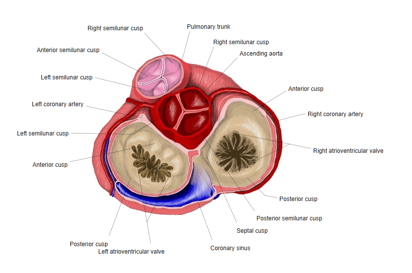
- Two atrioventricular valves: tricuspid (right) and mitral (left), held by chordae tendineae and papillary muscles.
- Two semilunar valves: pulmonary and aortic, at the outflow of the ventricles.
- All four valves are attached to the fibrous cardiac skeleton.
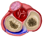
Base of the heart: the four valves seen from above
Wall of the heart
- Three layers: endocardium (inner lining), myocardium (muscle) and epicardium (= visceral layer of serous pericardium).
- The left ventricular wall is about three times thicker than the right, because it pumps against the higher systemic pressure.
Blood supply and innervation
- Arteries: right and left coronary arteries, the first branches of the ascending aorta.
- Veins: the cardiac veins, draining mostly into the coronary sinus.
- Nerves: the cardiac plexus (sympathetic fibres speed the heart; vagus nerve fibres slow it). The beat itself starts in the conducting system.
Heart from the front with the coronary arteries
Clinical notes
- Blockage of a coronary artery causes myocardial infarction (heart attack).
- The apex beat is normally felt in the left 5th intercostal space; it moves outward when the heart is enlarged.
References and sources
- Further reading: Heart, Wikipedia.