Latin: Ventriculus sinister
Overview
- The chamber that pumps oxygenated blood into the ascending aorta and so to the whole body.
- Forms the apex, most of the left border and the diaphragmatic surface of the heart; circular in cross section.
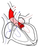
Chambers of the heart and the flow of blood
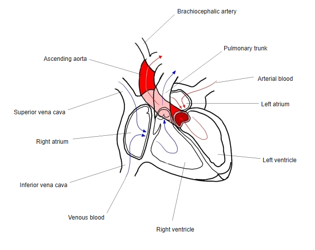
Internal features
- Inflow: through the mitral valve; outflow: through the aortic vestibule and the aortic valve.
- Two large papillary muscles (anterior and posterior), and fine trabeculae carneae.
- The wall is the thickest in the heart (about 10–15 mm), two to three times the right ventricle.
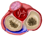
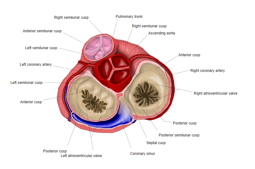
Base of the heart: the four valves seen from above
Clinical notes
- Long-standing high blood pressure or aortic stenosis thickens the left ventricular wall (left ventricular hypertrophy).
References and sources
- Further reading: Left ventricle, Wikipedia.