Latin: Atrium dextrum
Overview
- The chamber that receives deoxygenated blood returning from the body and from the heart wall.
- Forms the right border of the heart; passes blood to the right ventricle through the tricuspid valve.
Chambers of the heart and the flow of blood
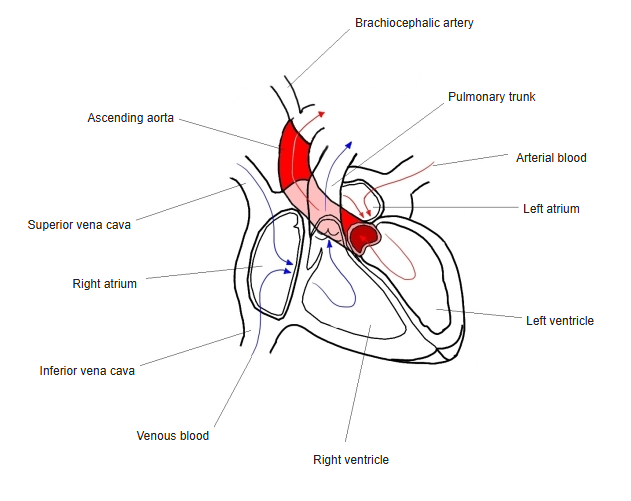
Openings
- Superior vena cava (no valve) and inferior vena cava (small valve of the inferior vena cava).
- Coronary sinus (valve of the coronary sinus), plus small openings of the smallest cardiac veins.
- Right atrioventricular orifice, guarded by the tricuspid valve.
Internal features
- Sinus venarum: smooth-walled posterior part where the venae cavae enter.
- Pectinate muscles: muscular ridges in the anterior wall and the right auricle.
- Crista terminalis: a ridge separating the two parts; outside it corresponds to the sulcus terminalis.
- On the septal wall: the fossa ovalis, remnant of the fetal foramen ovale.
- The sinoatrial node lies near the opening of the superior vena cava; the atrioventricular node lies near the opening of the coronary sinus.
Clinical notes
- Central venous catheters are placed with the tip near the junction of the superior vena cava and the right atrium.
References and sources
- Further reading: Right atrium, Wikipedia.