Latin: Venae thoracicae internae
Overview
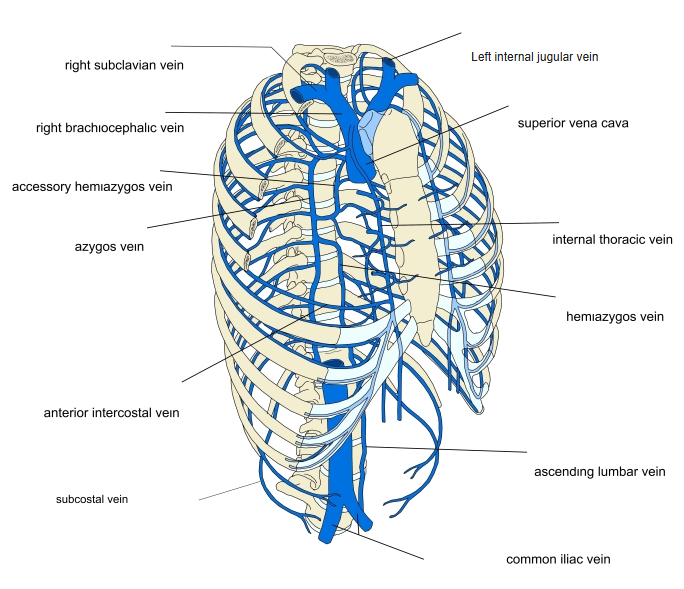
- Paired veins (often two per side below) that accompany the internal thoracic artery behind the costal cartilages, about 1 cm from the edge of the sternum.
- Begin by the union of the superior epigastric and musculophrenic veins.
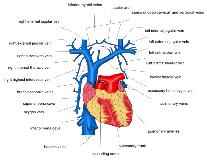
- Unite into one vein above the 3rd costal cartilage and end in the brachiocephalic vein.
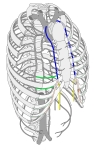
Internal thoracic veins
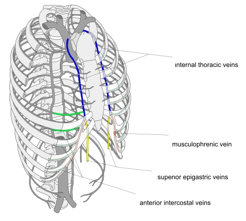
Tributaries and drainage
- Tributaries: anterior intercostal veins, perforating veins from the front of the chest wall and breast, pericardiacophrenic, mediastinal and thymic veins.
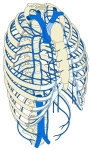
Veins of the thoracic wall 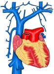
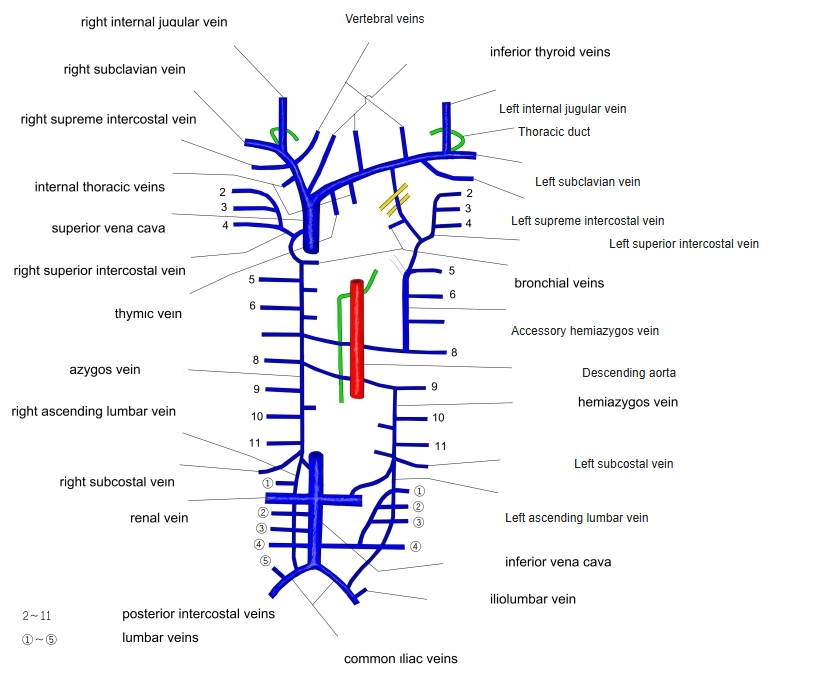
Superior vena cava and its tributaries 
Veins of the thorax (diagram)
Clinical notes
- Through the epigastric veins they connect the superior and inferior venae cavae, a route that enlarges when either vena cava is blocked.
References and sources
- Further reading: Internal thoracic vein, Wikipedia.