Latin: musculus coracobrachialis
Overview
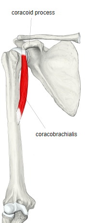
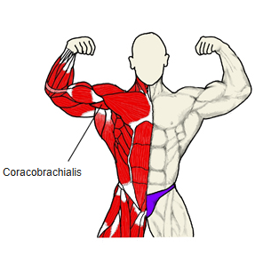
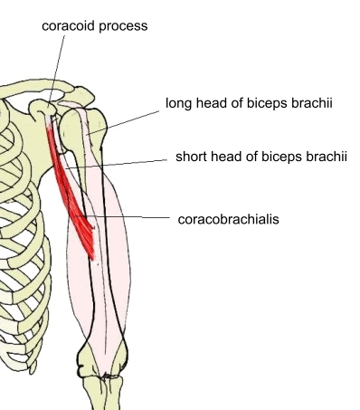
- A spindle-shaped muscle on the upper medial side of the arm, deep to the biceps brachii: one of the three flexors of the arm.
- The smallest of the three muscles attached to the coracoid process, with the pectoralis minor and the short head of the biceps brachii.
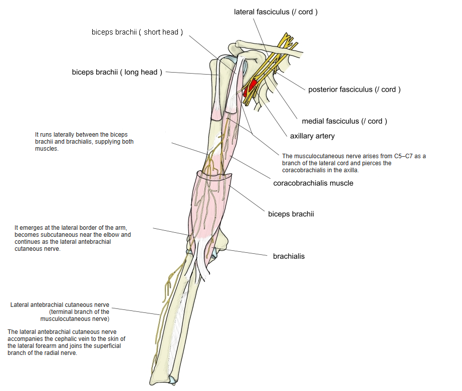
- Thick near its origin and thin near its insertion; pierced by the musculocutaneous nerve.
- Connected with the brachialis, the medial head of the triceps brachii, the biceps brachii and the pectoralis minor.
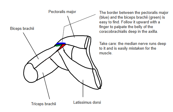
- Partly superficial in the axilla, so it can be palpated.
Position in the classification
Upper Limb › Arm › Flexor group
- Biceps brachii
- Coracobrachialis
- Brachialis
Illustrations
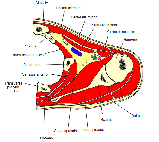
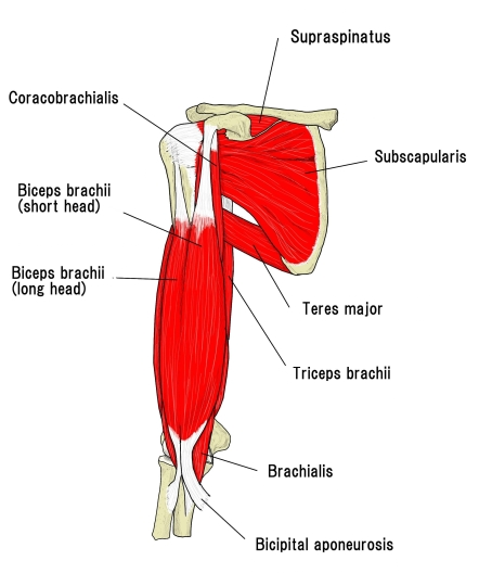
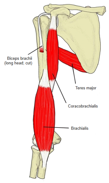
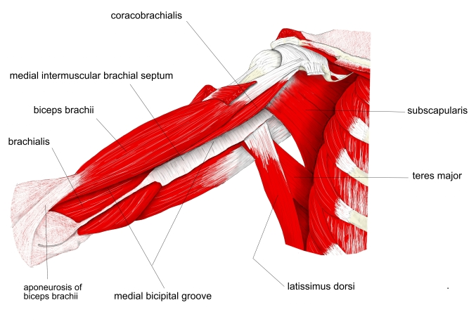
Origin
- Apex of the coracoid process of the scapula, together with the short head of the biceps brachii
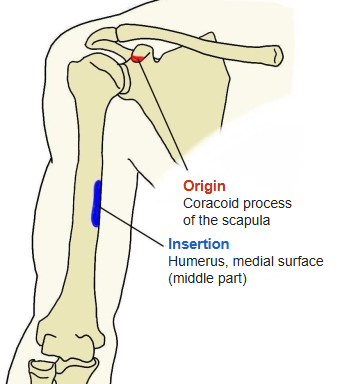
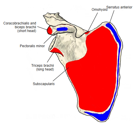
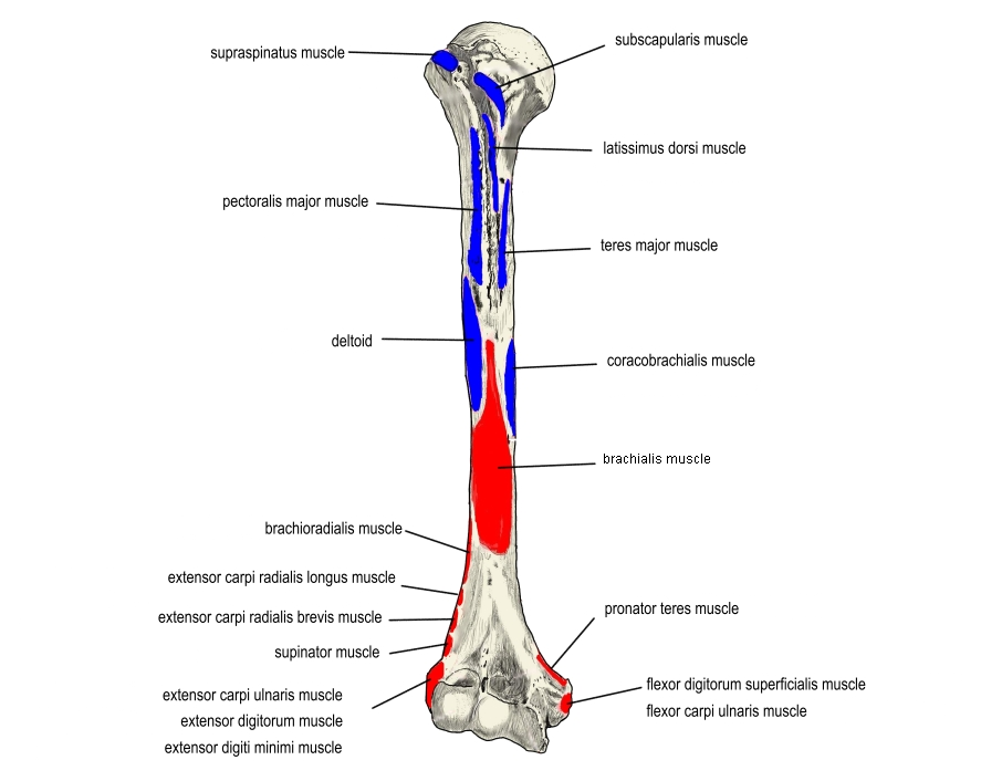
Insertion
- Middle of the medial surface of the humerus, distal to the crest of the lesser tubercle
- Between the attachments of the brachialis and the medial head of the triceps brachii
Action
- Flexion of the arm at the shoulder joint
- Adduction of the arm
Innervation
- Musculocutaneous nerve (C5–C7; some sources omit C5), which pierces the muscle
Blood supply
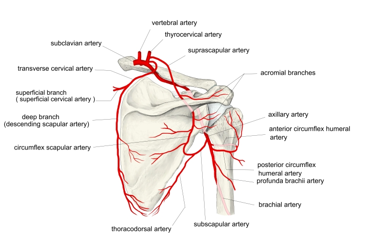
- Muscular branches of the brachial artery; some sources give the anterior and posterior circumflex humeral arteries
Anatomical variations
- May not be pierced by the musculocutaneous nerve
- Two or three separate parts may be present:
- Coracobrachialis brevis: from the base of the coracoid process to the shoulder joint capsule and the proximal humerus
- Coracobrachialis longus: from the coracoid process to the medial intermuscular septum of the arm, passing over the structures of the medial bicipital groove
Related terms
- Coracoid process
- Humerus
- Biceps brachii
- Brachialis
- Triceps brachii
- Pectoralis minor
- Musculocutaneous nerve
- Median nerve
- Anterior circumflex humeral artery
- Posterior circumflex humeral artery
References and sources
- Further reading: Coracobrachialis muscle, Wikipedia.