Latin: musculus digastricus
Overview
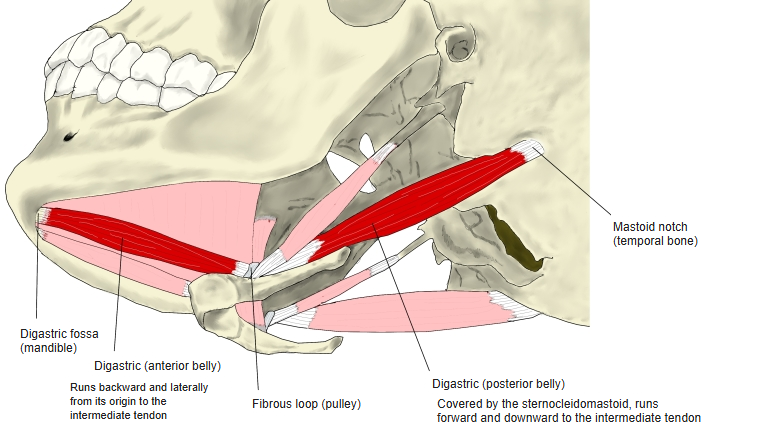
- A small muscle just below the mandible with two bellies joined by an intermediate tendon.
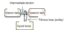
- The intermediate tendon passes through a fibrous sling (acting as a pulley) attached to the body and greater horn of the hyoid, and perforates the stylohyoid.
- The posterior belly is longer than the anterior one.
- The bellies come from different pharyngeal arches (anterior: 1st; posterior: 2nd) and so have different nerves.
- Other two-bellied muscles: the omohyoid and the occipitofrontalis. The stapedius also comes from the second pharyngeal arch; some textbooks describe it as split off from the posterior belly.
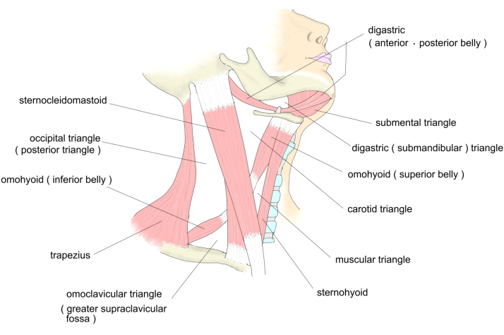
- With the mandible it bounds the submandibular (digastric) triangle.
Position in the classification
Neck › Superficial muscles of the neck › Suprahyoid muscles
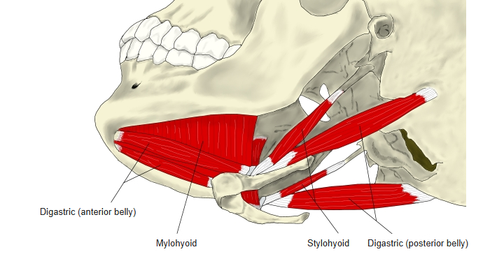
- Digastric
- Stylohyoid
- Mylohyoid
- Geniohyoid
Illustrations
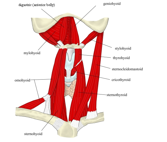
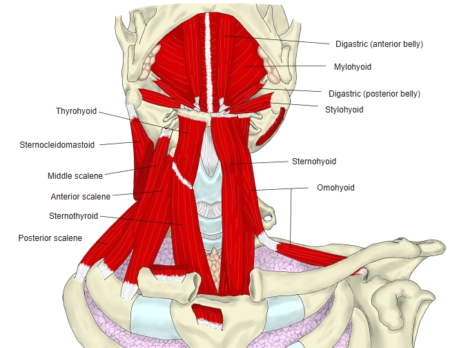
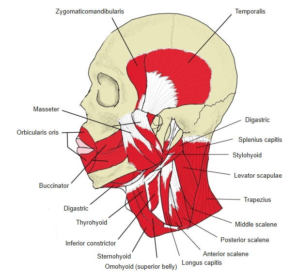
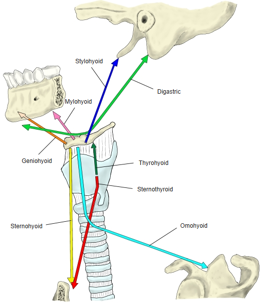
Origin / insertion
- Origin, anterior belly: digastric fossa of the mandible
- Origin, posterior belly: mastoid notch of the temporal bone
- Insertion: the intermediate tendon, held to the body and greater horn of the hyoid by the fibrous loop
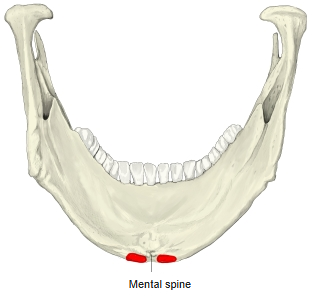
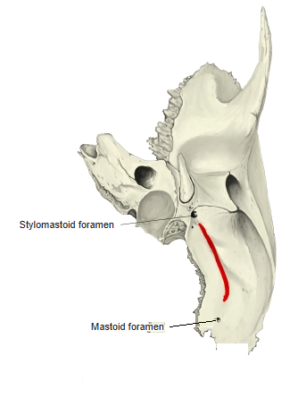
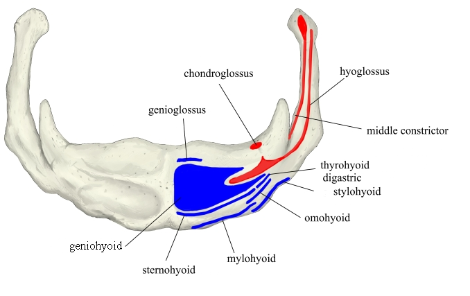
The attachment areas on the temporal bone are only approximate.
Action
- Raises the hyoid bone and with it the base of the tongue (swallowing)
- With the hyoid fixed, depresses the mandible (opens the mouth; helps chewing)
Innervation
- Anterior belly: nerve to mylohyoid (inferior alveolar nerve, mandibular nerve V3)
- Posterior belly: digastric branch of the facial nerve
Blood supply
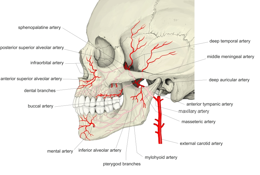
- Artery: anterior belly – submental branch of the facial artery and the mylohyoid branch of the inferior alveolar artery; posterior belly – occipital artery
- Vein: not described in the sources used here
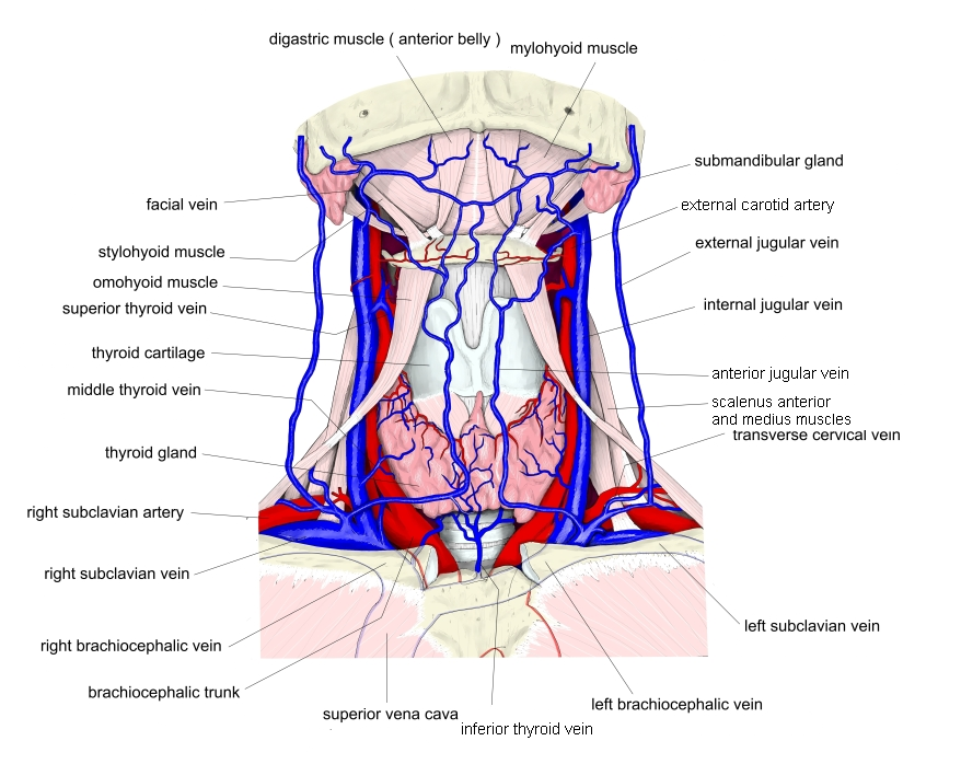
Related terms
- Mylohyoid
- Stylohyoid
- Geniohyoid
- Omohyoid
- Stapedius
- Hyoid bone
References and sources
- Further reading: Digastric muscle, Wikipedia.