Latin: musculus obliquus externus abdominis
Overview
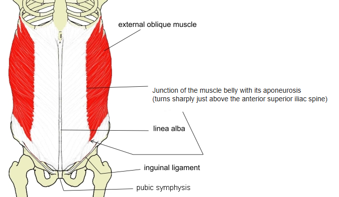
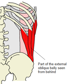
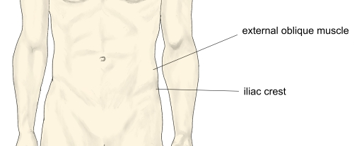
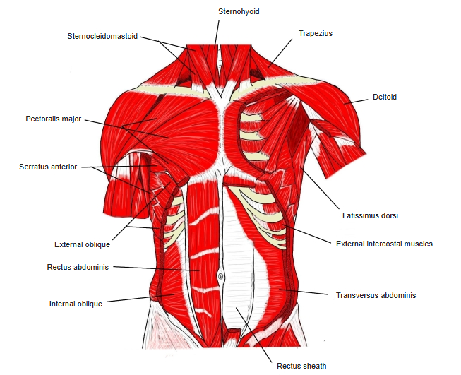
- The largest and outermost of the three flat muscles of the lateral abdominal wall; it corresponds to the external intercostals of the thorax.
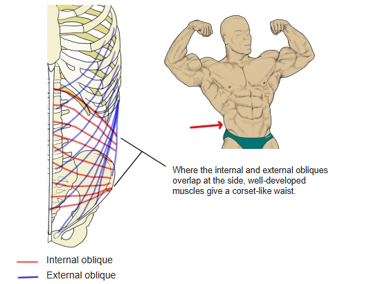
- Its fibres run obliquely downward and forward over the side of the trunk.
- The upper digitations interlock with the serratus anterior, the lower ones with the latissimus dorsi.
- Its aponeurosis contributes to the anterior layer of the rectus sheath, meets the other side at the linea alba and forms the inguinal ligament.
- Between the latissimus dorsi, the external oblique and the iliac crest lies the inferior lumbar triangle (of Petit).
Position in the classification
Trunk › Abdomen › Lateral abdominal muscles
- External oblique
- Internal oblique
- Transversus abdominis
Illustrations
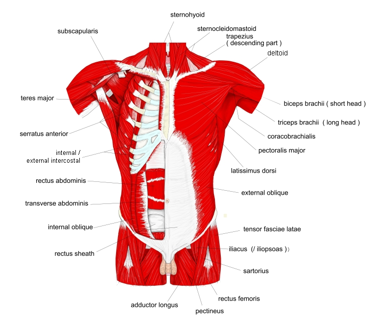
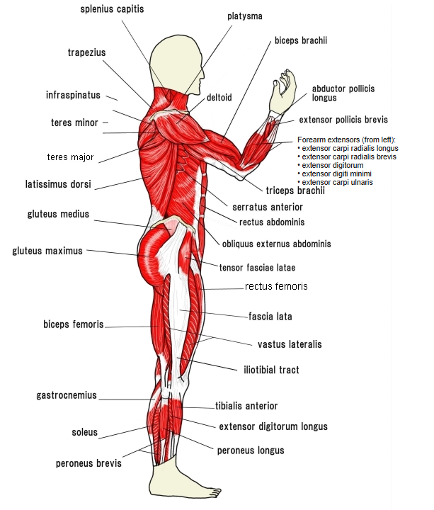
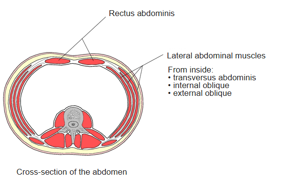
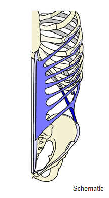
Origin / insertion
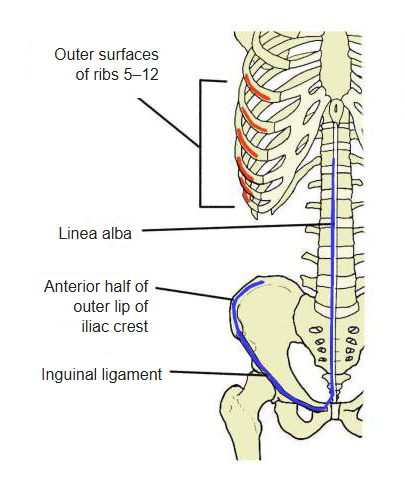
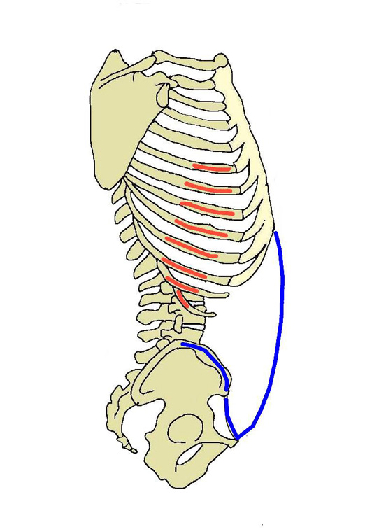
- Origin: outer surfaces of ribs 5–12 (eight digitations)
- Insertion: anterior half of the outer lip of the iliac crest; through its aponeurosis into the linea alba and the pubic tubercle and crest (its free lower border forms the inguinal ligament)
Action
- Both sides: flex the trunk
- One side: bends the trunk to the same side and rotates it to the opposite side
- Depresses the thorax and tilts the pelvis backward
- Compresses the abdomen (raises intra-abdominal pressure)
Innervation
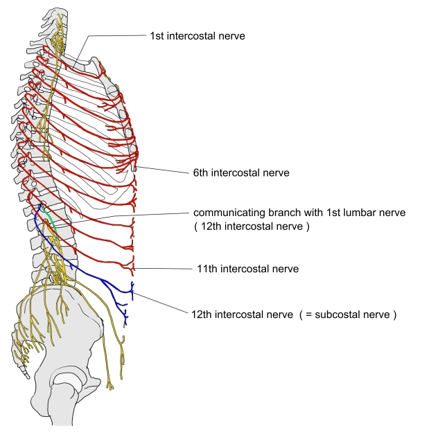
- Lower intercostal nerves and the subcostal nerve (T7–T12; some sources T5–T12); some sources add branches of the iliohypogastric and ilioinguinal nerves
Blood supply
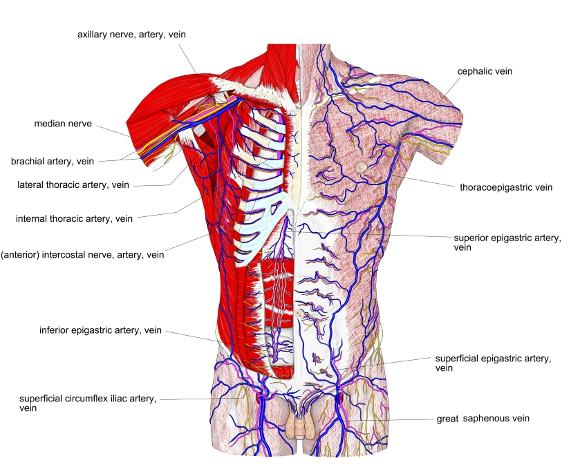
- Muscular branches of the superior and inferior epigastric arteries
- Lower posterior intercostal and subcostal arteries (upper part); deep circumflex iliac or iliolumbar arteries (lower part)
Related terms
- Internal oblique
- Transversus abdominis
- Rectus abdominis
- Serratus anterior
- Latissimus dorsi
- Inguinal ligament
- Linea alba
References and sources
- Further reading: Abdominal external oblique muscle, Wikipedia.
- Gray H. Anatomy of the Human Body, 20th ed. (1918), “6d. The Muscles and Fasciæ of the Abdomen”, Bartleby.com.