Latin: musculus geniohyoideus
Overview
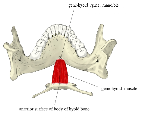
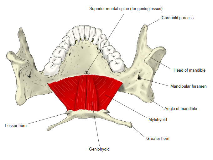
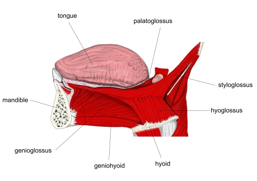
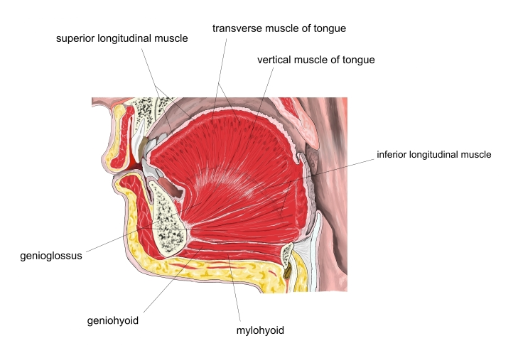
- A narrow suprahyoid muscle lying above the mylohyoid, next to the midline.
- Paired: the left and right muscles lie side by side.
- Runs from the chin backward and slightly downward to the hyoid bone.
Position in the classification
Neck › Superficial muscles of the neck › Suprahyoid muscles
- Digastric
- Stylohyoid
- Mylohyoid
- Geniohyoid
Illustrations
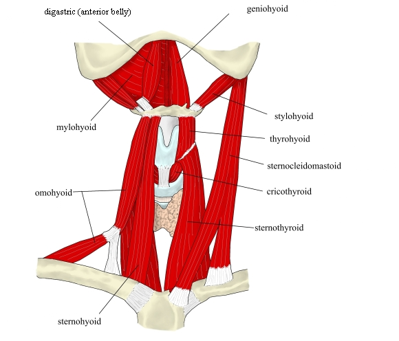
Origin / insertion
- Origin: inferior mental spine on the posterior surface of the mandibular symphysis
- Insertion: anterior surface of the body of the hyoid bone
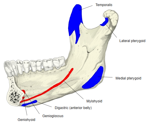
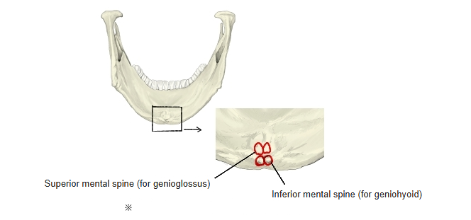
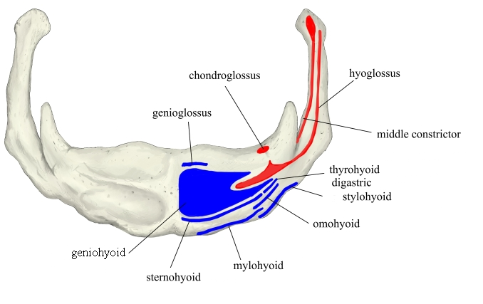
Action
- Pulls the hyoid bone forward and upward (e.g. in swallowing), widening the upper airway
- With the hyoid fixed, depresses the mandible
Innervation
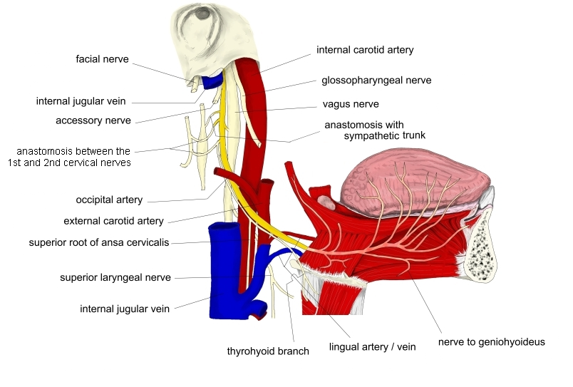
- Fibres of the first cervical nerve (C1), carried by the hypoglossal nerve
Blood supply
- Branches of the lingual artery
Compiled from standard anatomy references.
Related terms
- Suprahyoid muscles
- Mylohyoid
- Digastric
- Stylohyoid
- Genioglossus
- Inferior mental spine
- Hypoglossal nerve
References and sources
- Further reading: Geniohyoid muscle, Wikipedia.