Latin: musculus flexor digitorum brevis
Overview
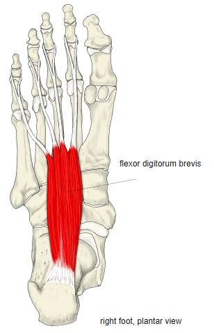
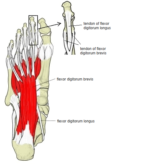
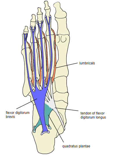
- Lies in the middle of the sole, covered by the plantar aponeurosis; the quadratus plantae and the tendons of the flexor digitorum longus lie deep to it.
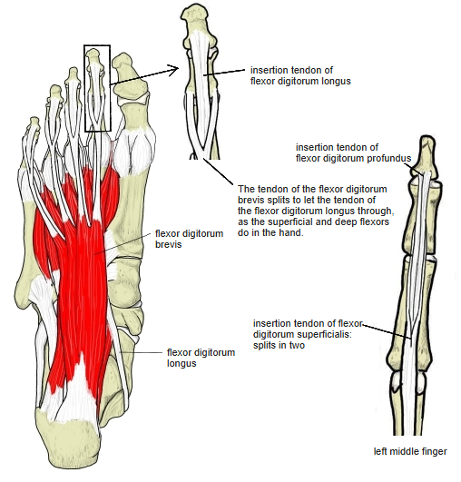
- Corresponds to the flexor digitorum superficialis of the hand: each tendon splits in two to let the tendon of the flexor digitorum longus through.
- At the toes the tendons are enclosed in synovial sheaths, which begin only at the distal quarter of the metatarsals.
- The tendon to the 5th toe is sometimes missing.
Position in the classification
Lower Limb › Foot › Sole of foot › Metatarsal muscles
- Flexor digitorum brevis
- Quadratus plantae
- Lumbricals (foot)
- Dorsal interossei (foot)
- Plantar interossei
Illustrations
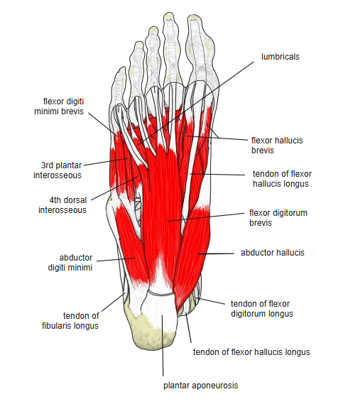
Origin / insertion
- Origin:
- Medial process of the calcaneal tuberosity, the central part of the plantar aponeurosis and the adjacent intermuscular septa
- Also given: under surface of the calcaneal tuberosity and part of the plantar ligaments near the calcaneus
- Insertion: sides of the middle phalanges of the 2nd–5th toes (each tendon divides around a tendon of the flexor digitorum longus)
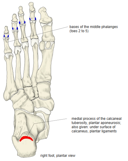
Action
- Flexes the middle phalanges of the 2nd–5th toes
- When the foot bears weight, helps to support the medial and lateral longitudinal arches
Innervation
- Medial plantar nerve (L5, S1)
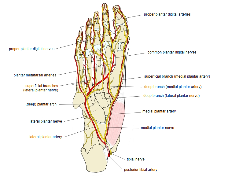
Blood supply
- Medial and lateral plantar arteries
Related terms
- Flexor digitorum longus
- Quadratus plantae
- Flexor digitorum superficialis
- Plantar aponeurosis
- Calcaneus
- Medial plantar nerve
References and sources
- Further reading: Flexor digitorum brevis muscle, Wikipedia.