Latin: musculus pterygoideus lateralis (also called the external pterygoid)
Overview
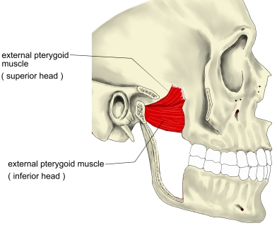
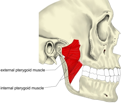
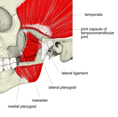
- A muscle of mastication with two heads (superior and inferior), lying above the medial pterygoid.
- The only muscle of mastication that helps to open the mouth.
- Variation: the upper head may be separate or joined to the temporalis.
Position in the classification
Head › Muscles of mastication
- Masseter
- Temporalis
- External pterygoid
- Medial pterygoid
Illustrations
Origin / insertion
- Origin, superior head: infratemporal crest and infratemporal surface of the greater wing of the sphenoid
- Origin, inferior head: lateral surface of the lateral pterygoid plate (some sources also include the maxillary tuberosity)
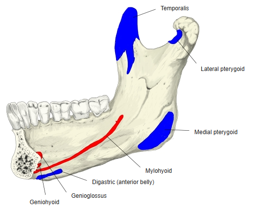
- Insertion: pterygoid fovea on the front of the neck of the condylar process of the mandible; capsule and front margin of the articular disc of the temporomandibular joint
Action
- Pulls the head of the mandible forward
- Both sides: protrude the mandible and open the mouth
- One side: moves the chin to the opposite side (grinding), usually with the medial pterygoid
Innervation
- Lateral pterygoid nerve, from the anterior division of the mandibular nerve (third division of the trigeminal nerve)
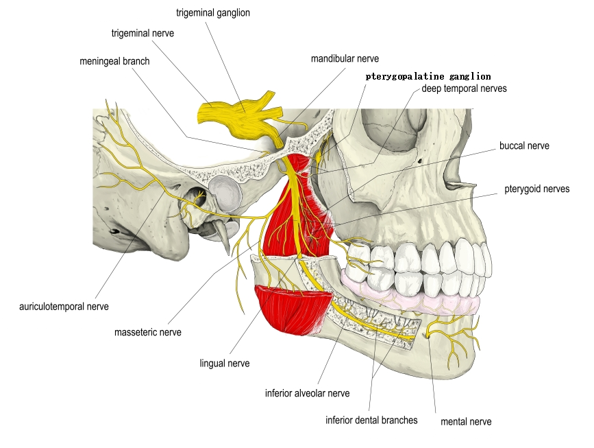
Blood supply
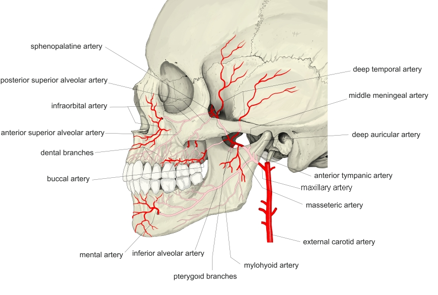
- Artery: pterygoid branches of the maxillary artery (a terminal branch of the external carotid artery)
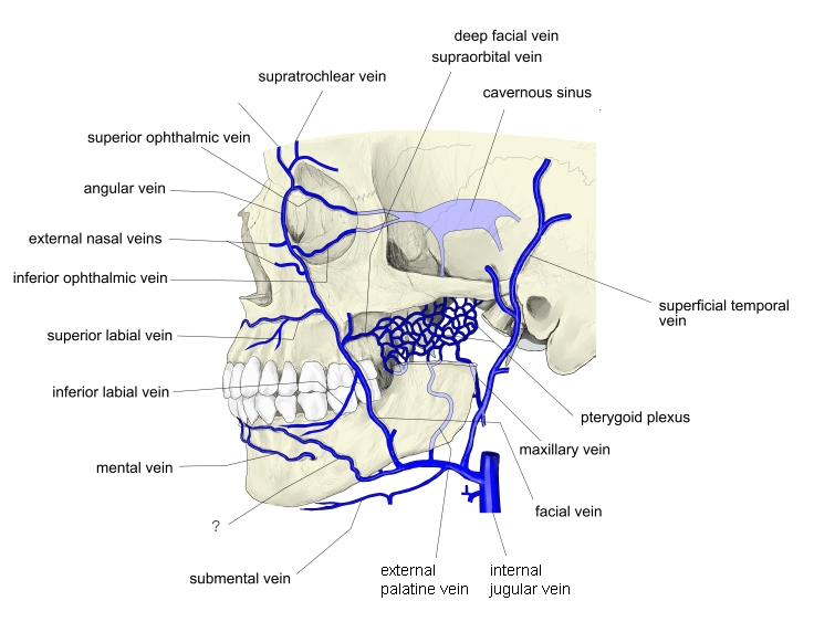
- Vein: pterygoid venous plexus (its tributaries correspond to the branches of the maxillary artery)
Related terms
- Muscles of mastication
- Medial pterygoid
- Temporalis
- Masseter
- Mandibular nerve
- Maxillary artery
References and sources
- Further reading: Lateral pterygoid muscle, Wikipedia.