Latin: nervus trigeminus
Overview
- The fifth cranial nerve (V), the largest cranial nerve; the nerve of the first pharyngeal arch.
- A mixed nerve: a large sensory root for the face, scalp, eye, nose, mouth and teeth, and a small motor root for the muscles of mastication.
- The sensory root has far more fibres (about 140,000) than the motor root (about 9,000).
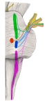
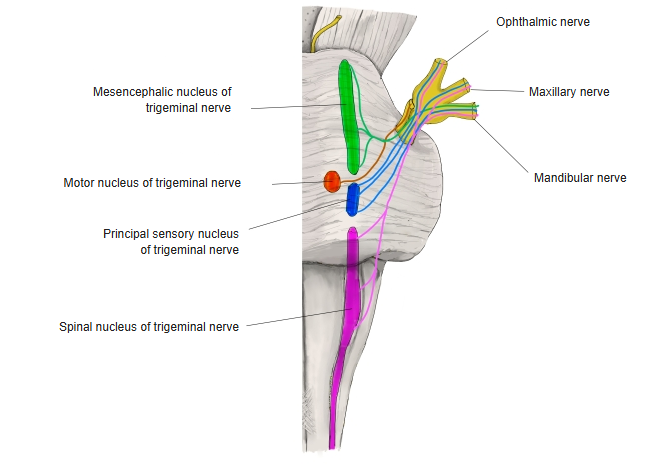
Nuclei of the trigeminal nerve 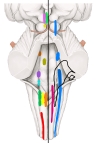
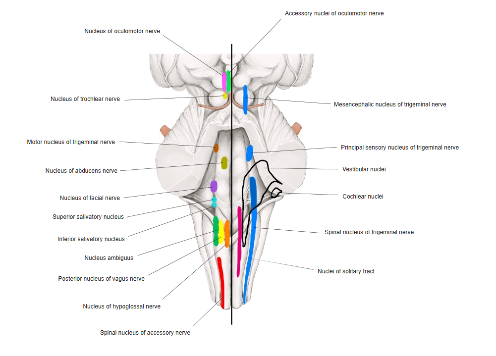
Cranial nerve nuclei in the brainstem
Nuclei
| Nucleus | Position | Function |
|---|---|---|
| Mesencephalic nucleus | Midbrain | Proprioception from the muscles of mastication and the teeth; its cells are like those of a spinal ganglion |
| Principal (chief) sensory nucleus | Pons, lateral to the motor nucleus | Touch and pressure from the head and face |
| Spinal nucleus | From the pons down to the upper cervical cord | Pain and temperature (mainly in its caudal part) |
| Motor nucleus | Pons, medial to the principal sensory nucleus | Muscles of mastication; reflex control of biting force via the mesencephalic nucleus |
Trigeminal ganglion
- The sensory (semilunar, Gasserian) ganglion in the trigeminal impression near the apex of the petrous part of the temporal bone.
- Its neurons are arranged by area: ophthalmic cells medially, mandibular laterally and maxillary between them.
- The three divisions leave the ganglion and the skull at once.
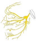
Trigeminal nerve and its branches (schematic)
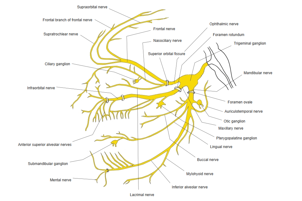
Branches
| Division | Leaves the skull through | Type |
|---|---|---|
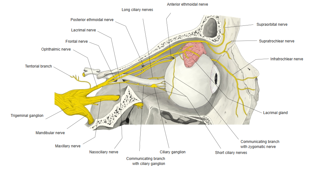
| Ophthalmic nerve (V1) | Superior orbital fissure | Sensory |
| Maxillary nerve (V2) | Foramen rotundum | Sensory |
| Mandibular nerve (V3) | Foramen ovale | Sensory and motor (with the motor root) |
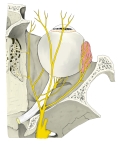
Branches of the ophthalmic nerve 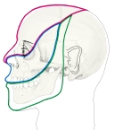
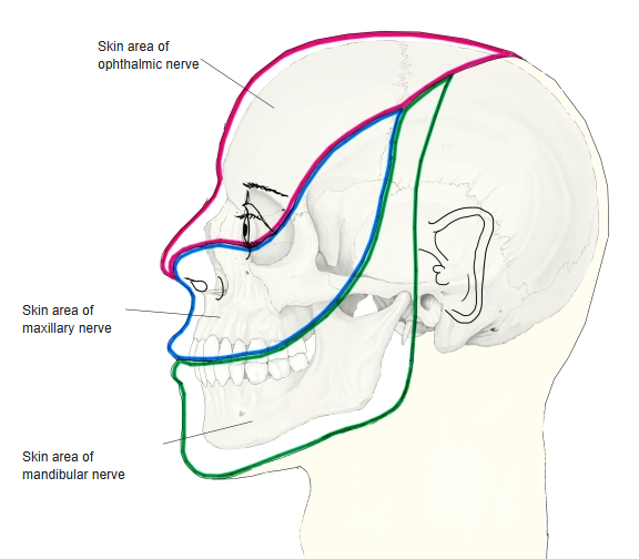
Skin areas of the trigeminal nerve
References and sources
- Further reading: Trigeminal nerve, Wikipedia.