Latin: musculus obturatorius externus
Overview
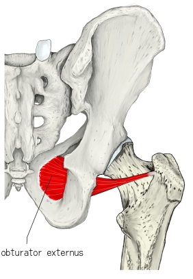
- A flat, triangular muscle covering the outer surface of the obturator membrane.
- Its tendon runs behind the neck of the femur; it may be hidden by the quadratus femoris.
- Both obturator muscles are about 1 cm thick near the obturator membrane.
- Some atlases (e.g. Prometheus) count it among the adductors; others place it with the outer hip muscles.
- Variation: the obturator nerve and vessels often divide it into two parts.
Position in the classification
Lower Limb › Hip › Outer hip muscles › Deep lateral rotators
Illustrations
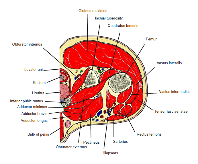
Origin / insertion
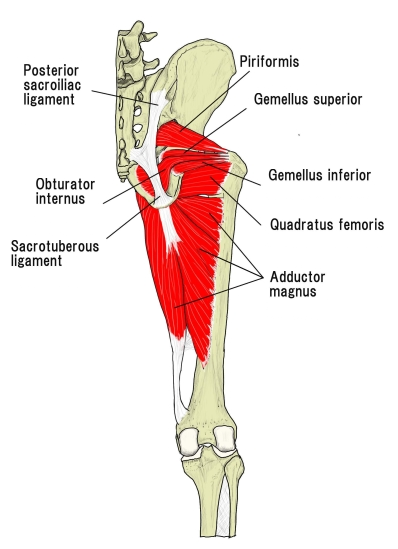
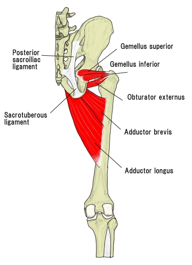
- Origin: outer surface of the hip bone around the obturator foramen; outer surface of the obturator membrane
- Insertion: trochanteric fossa of the femur
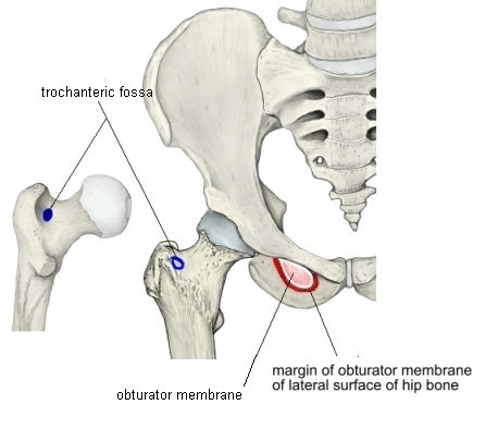
Action
- Rotates the thigh laterally
- Helps to raise and adduct the thigh
- Stabilises the hip joint
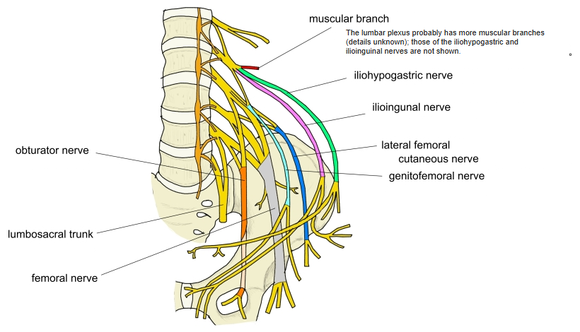
Innervation
- Obturator nerve (L3, L4)
Blood supply
- Obturator artery
- Medial circumflex femoral artery
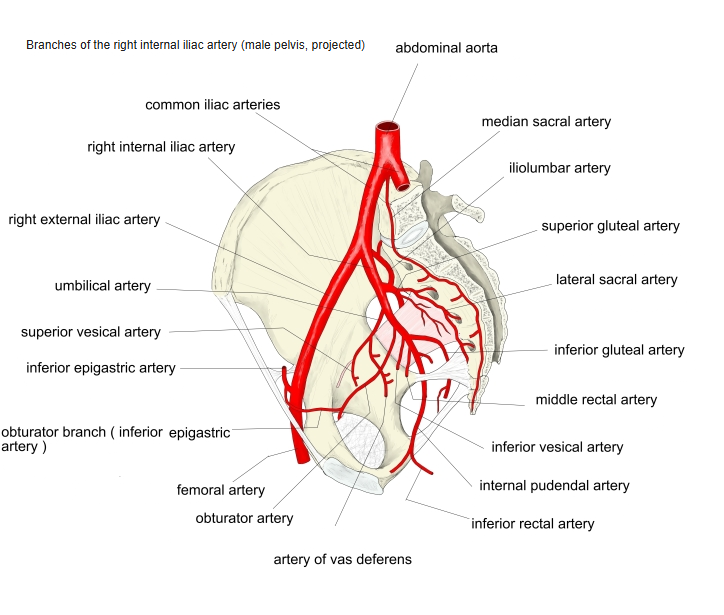
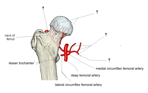
The figure of the medial circumflex femoral artery was drawn from several sources and may not be exact.
Related terms
- Obturator internus
- Quadratus femoris
- Adductor brevis
- Obturator nerve
- Medial circumflex femoral artery
References and sources
- Further reading: External obturator muscle, Wikipedia.