Latin: musculus splenius cervicis · also called the splenius colli
Overview
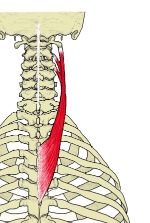
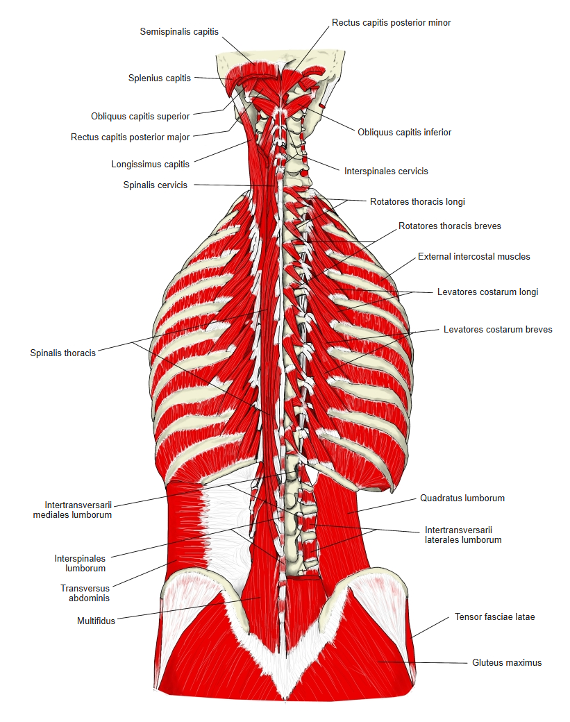
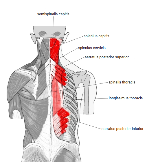
- A thin, flat muscle in the posterior neck and upper back, deep to the trapezius, rhomboids, levator scapulae and serratus posterior superior.
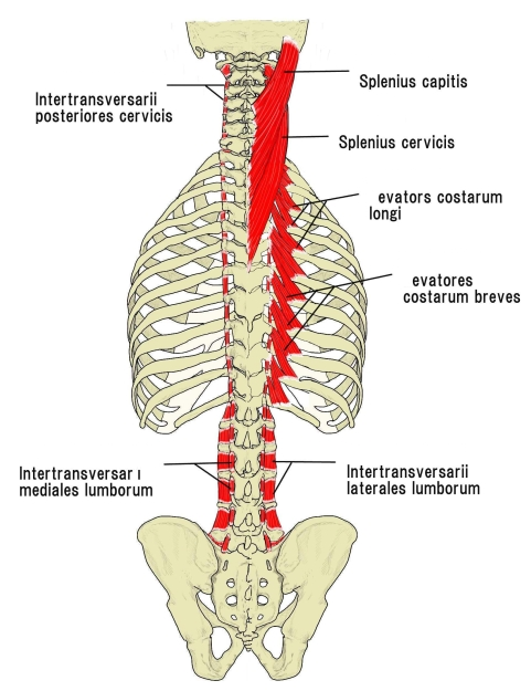
- Forms the splenius group with the splenius capitis, which lies above and medial to it; the two are often partly fused.
- Broad at its thoracic origin, it narrows into two (sometimes three) tendinous slips on the upper cervical vertebrae.
- Does not attach to the skull.
Position in the classification
Trunk › Back › Deep back muscles › Second layer › Intrinsic back muscles › Splenius muscles
- Splenius capitis
- Splenius cervicis
Illustrations
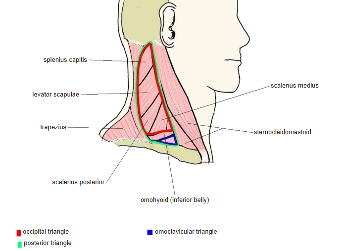
Origin
- Spinous processes of T3–T6 (upper limit T3 or T4, depending on the source)
Insertion
- Posterior tubercles of the transverse processes of C1–C2
- Inserts next to the levator scapulae, which also attaches to the upper cervical transverse processes.
- An extra slip to C3 may occur (see Variations).
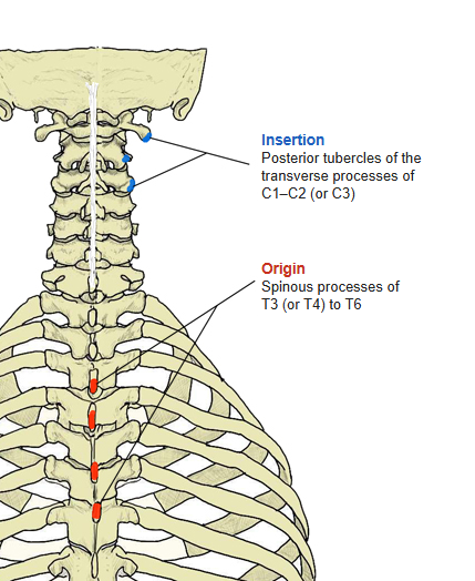
Action
- Bilateral: extends the cervical spine
- Unilateral: rotates the neck to the same side
- Unilateral: assists lateral flexion of the neck to the same side
No attachment to the skull — it moves the head only indirectly, through the cervical spine.
Innervation
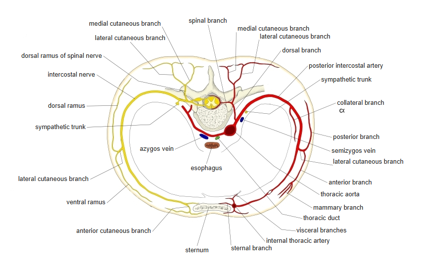
- Lateral branches of the posterior (dorsal) rami of the cervical spinal nerves
- Levels differ between sources: C2–C5, or C1–C4 (occasionally C5, rarely C6)
Blood supply
- Muscular branches of the descending branch of the occipital artery
- Superficial branch of the transverse cervical artery
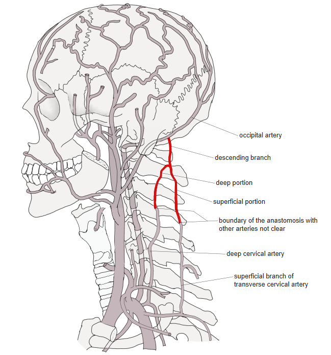
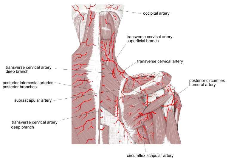
The boundary between the descending branch and its anastomoses with neighbouring arteries is not clearly defined.
Anatomical variations
- May be absent
- May be completely separate from the splenius capitis
- Extra insertion slip to the transverse process of C3
- Accessory muscle (rhomboatlanticus, also called splenius accessorius; about 8% in classic reports): lowest cervical or highest thoracic spinous processes → transverse process of the atlas
Related terms
- Splenius capitis
- Splenius muscles
- Intrinsic back muscles
- Erector spinae
- Transversospinales
- Suboccipital muscles
- Levator scapulae
- Serratus posterior superior
- Spinous process
- Transverse process
- Cervical nerves
- Occipital artery
- Transverse cervical artery
References and sources
- Further reading: Splenius cervicis muscle, Wikipedia.