Latin: nervi spinales
Overview
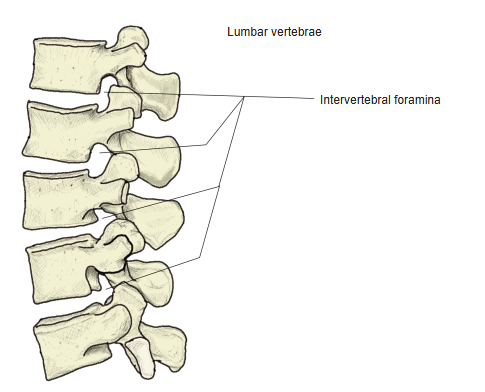
- 31 pairs of nerves that leave the spinal cord and pass out of the vertebral canal through the intervertebral foramina.
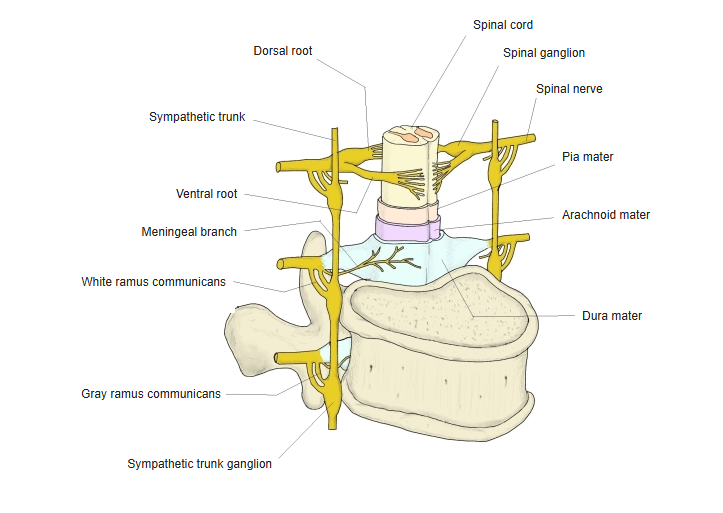
- Each nerve is formed by a ventral (anterior) root and a dorsal (posterior) root, so it is a mixed nerve.
- Besides somatic motor and sensory fibres, they carry sympathetic fibres (thoracic and upper lumbar segments) and parasympathetic fibres (sacral segments).
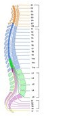
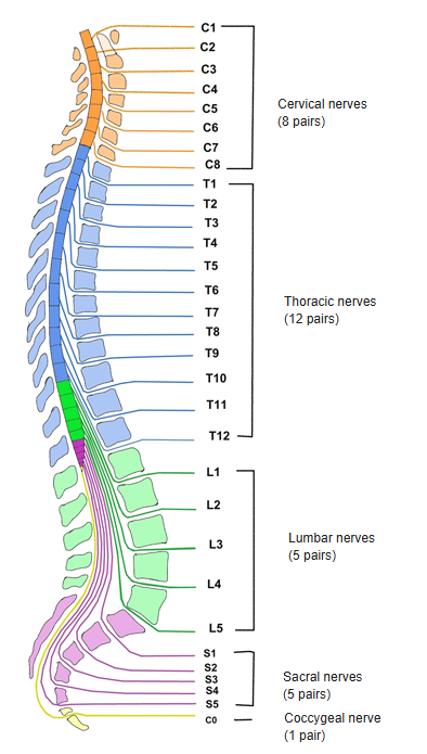
The 31 pairs of spinal nerves 
Intervertebral foramina (lumbar)
Divisions
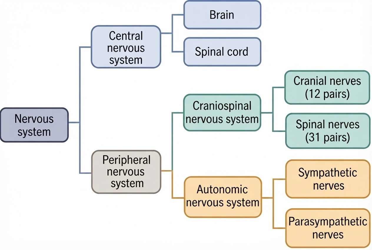
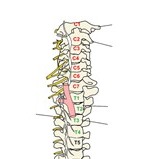
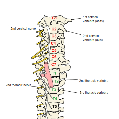
- Cervical nerves are numbered after the vertebra below them (C2 passes between C1 and C2); all others after the vertebra above them (T2 passes between T2 and T3).
- C8 has no vertebra of its own: it leaves between C7 and T1.
| Group | Pairs | Nerves |
|---|---|---|
| Cervical nerves | 8 | C1–C8 |
| Thoracic nerves | 12 | T1–T12 |
| Lumbar nerves | 5 | L1–L5 |
| Sacral nerves | 5 | S1–S5 |
| Coccygeal nerve | 1 | Co |
Cervical and upper thoracic nerves 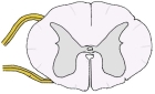
Spinal cord in cross-section
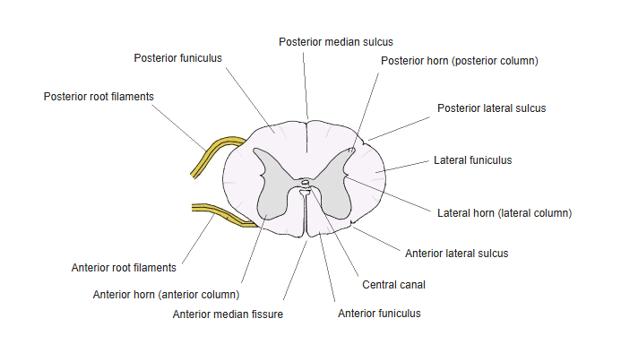
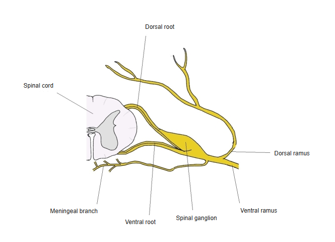
Ventral and dorsal roots
- Ventral root: motor fibres to skeletal muscle; preganglionic sympathetic fibres (thoracolumbar) and parasympathetic fibres (sacral).
- Dorsal root: sensory fibres, mainly from the skin. It carries the spinal (dorsal root) ganglion, which holds the cell bodies of the sensory neurons; there are no synapses in it.
Components of a spinal nerve 
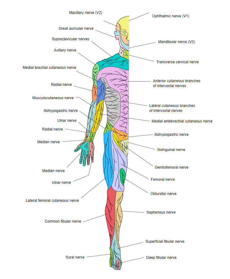
Cutaneous nerves of the body (front) 
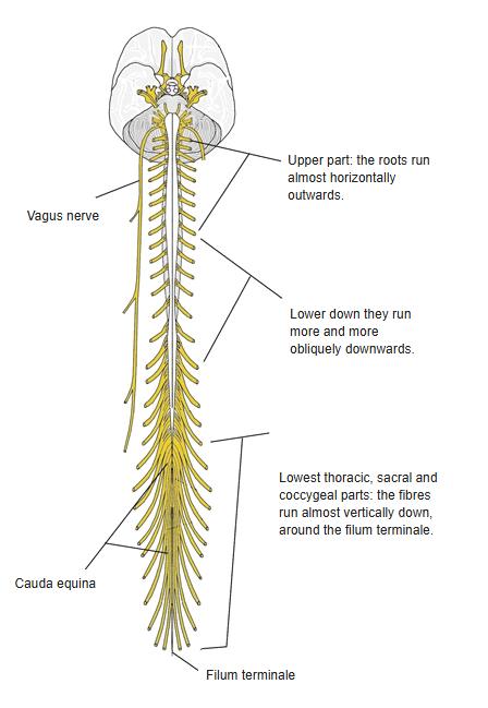
Cauda equina
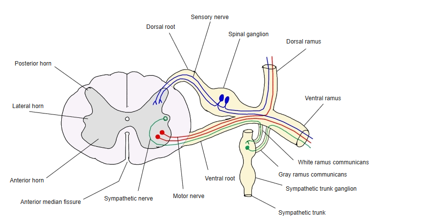
Ventral and dorsal rami
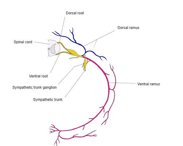
- Just outside the foramen each nerve divides into a ventral ramus and a dorsal ramus.
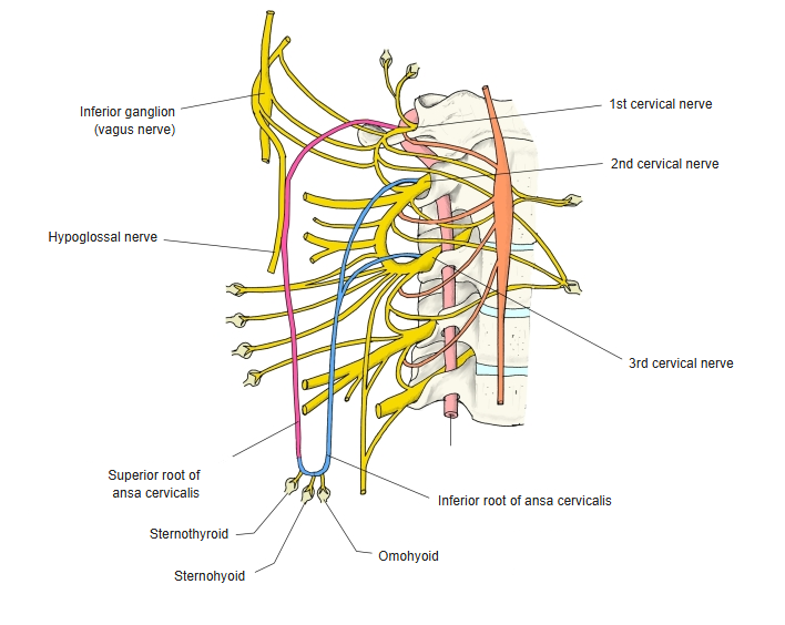
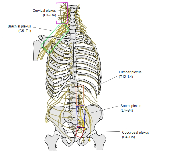
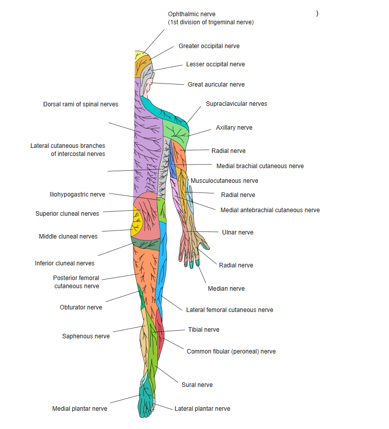
- Ventral rami: the lateral and front parts of the trunk and the limbs. Except in the thorax they join to form the plexuses: cervical (C1–C4), brachial (C5–T1), lumbar (T12–L4), sacral (L4–S4), pudendal (S2–S4) and coccygeal (S4–Co).
- Dorsal rami: the deep (intrinsic) muscles of the back and the skin of the back. The dorsal rami of C1 and C2 are larger than their ventral rami: the suboccipital nerve and the greater occipital nerve.
Roots and rami of a spinal nerve 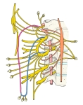
Ansa cervicalis 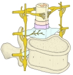
Spinal nerve and the vertebral column 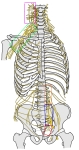
Plexuses of the spinal nerves
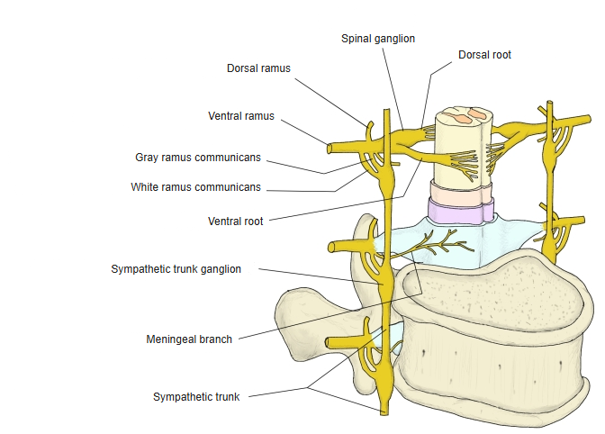
Cauda equina and communicating branches
- Cauda equina: the roots of the lower nerves run almost straight down in the vertebral canal below the end of the spinal cord, forming a bundle around the filum terminale.
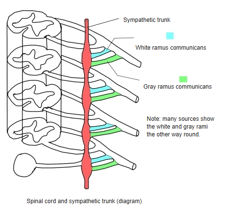
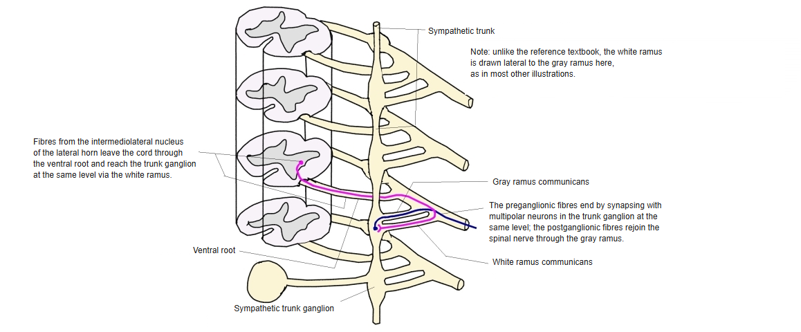
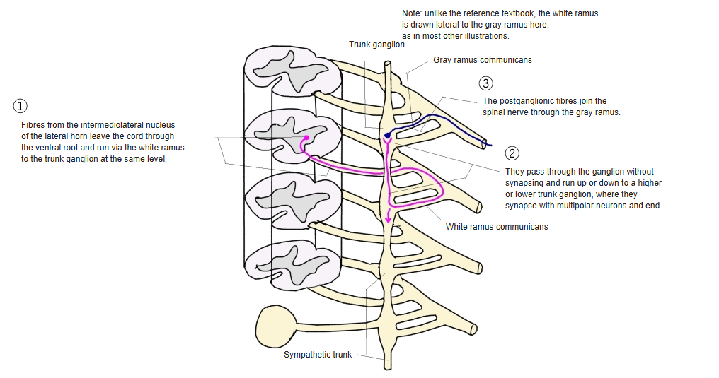
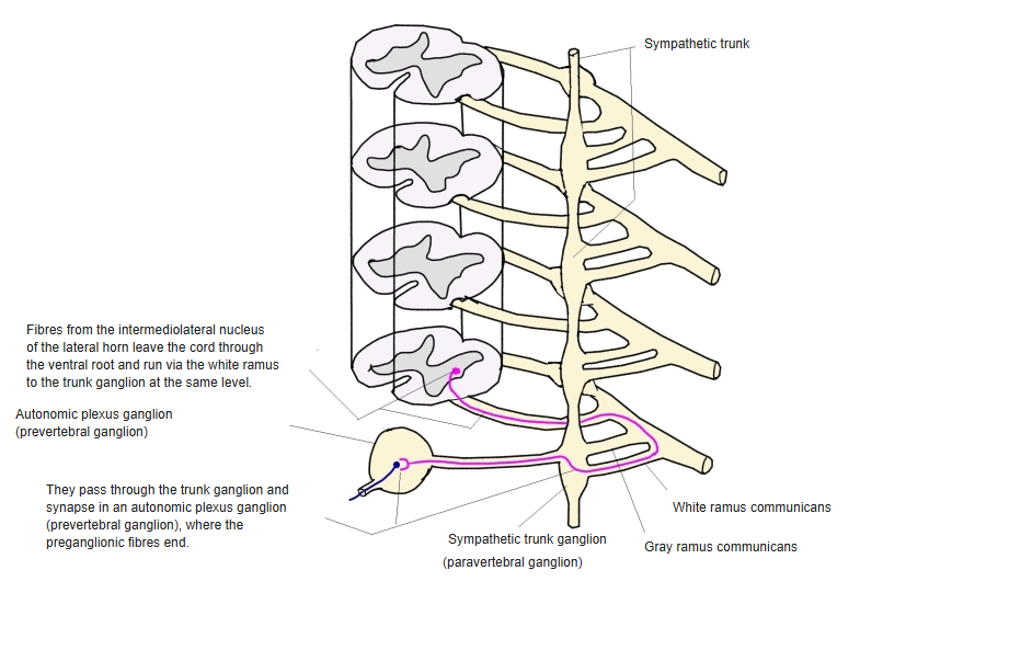
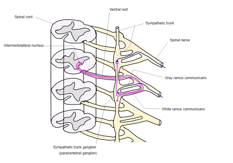
- White ramus communicans: from the spinal nerve to a ganglion of the sympathetic trunk; myelinated preganglionic sympathetic fibres from the intermediolateral nucleus (T1–L2).
- A preganglionic fibre may synapse in the ganglion at the same level, run up or down to a ganglion at another level, or pass through to a prevertebral ganglion.
- Grey ramus communicans: mostly unmyelinated postganglionic fibres returning from the sympathetic ganglion to every spinal nerve.
- Meningeal branch: a small recurrent branch back through the intervertebral foramen to the meninges.
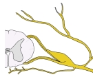
Meningeal branch of a spinal nerve 
Cutaneous nerves of the whole body (back) 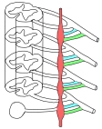
White and gray rami communicantes 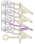
Sympathetic fibres: synapse at the same level 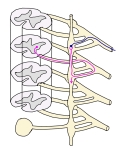
Sympathetic fibres: synapse at another level 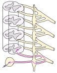
Sympathetic fibres: synapse in a prevertebral ganglion 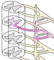
Intermediolateral nucleus and rami communicantes 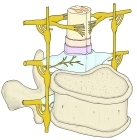
Spinal nerve, meninges and meningeal branch
References and sources
- Further reading: Spinal nerves, Wikipedia.