Inferior horn of lateral ventricle (Latin: cornu inferius ventriculi lateralis)
Also called the temporal horn
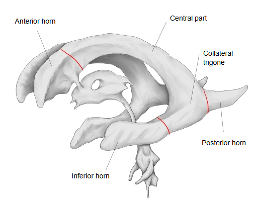
The inferior horn of the lateral ventricle is:
The main parts of the lateral ventricle are as follows.
ventricle
- Anterior horn : lies in the frontal lobe
- Central part : lies in the parietal lobe
- Inferior horn : lies in the temporal lobe and ends a short distance behind its front end; the largest horn
- Posterior horn : lies in the occipital lobe
Structure
The walls of the inferior horn are made up as follows.
· Floor (medial part): the hippocampus, which ends in front as the pes hippocampi, with a few rounded elevations (digitations); lateral to it lies the collateral eminence, and along its medial edge run the fimbria of the hippocampus and the choroidal fissure.
· Roof: the tapetum, the tail of the caudate nucleus, the stria terminalis and the line of attachment of the choroid plexus (taenia choroidea).
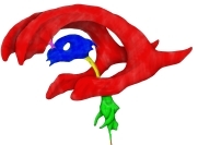 Ventricles: left side (coloured)
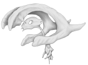 Ventricles: left side
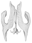 Ventricles: from above
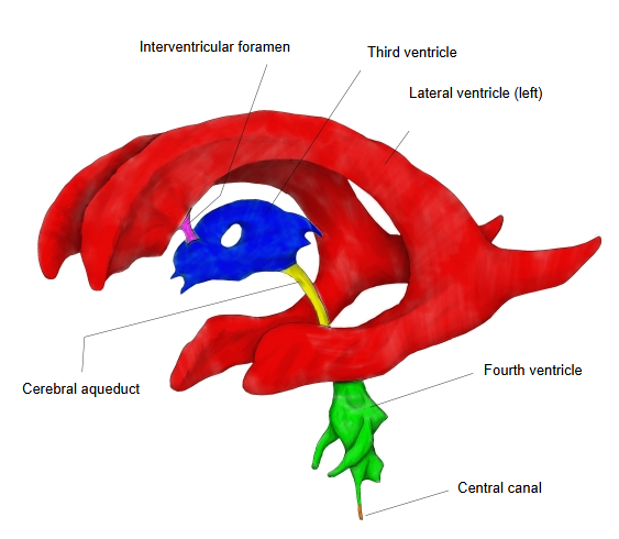
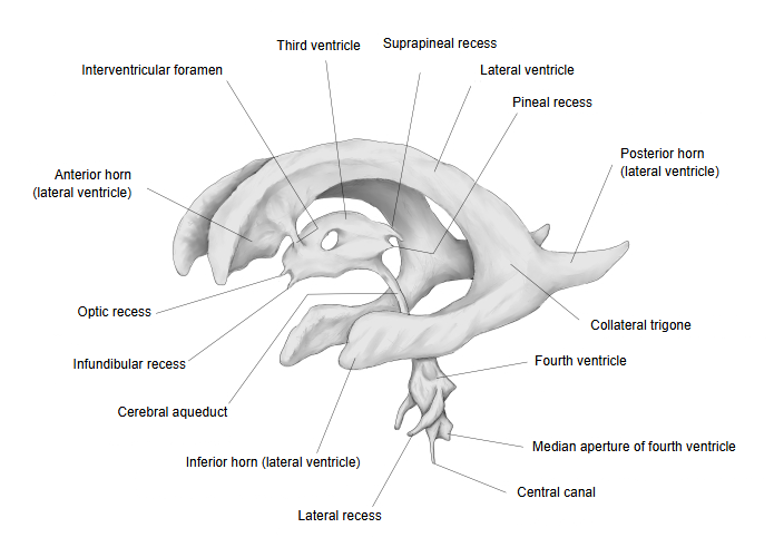
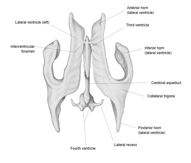
The inferior horn projects forwards and downwards into the temporal lobe. It begins at the atrium (where the collateral trigone lies in the floor), where it meets the central part and the posterior horn, and curves round the back of the thalamus, first backwards, outwards and downwards, then forwards; it ends about 2.5 cm from the temporal pole, and its direction roughly follows the superior temporal sulcus on the brain surface. Its roof and lateral wall are formed mainly by the tapetum, fibres of the corpus callosum that spread out from its splenium; the tail of the caudate nucleus and the stria terminalis run forwards along its medial part. On the floor, the collateral sulcus raises the collateral eminence, which widens behind into the triangular collateral trigone and continues to the posterior horn. The floor and medial wall also bear the hippocampus and the fimbria of the hippocampus, the continuation of the crus of the fornix. The choroid plexus of the lateral ventricle, covered by ependyma, enters along the medial side through the lower part of the choroidal fissure.
Reference websites
[Wikipedia]
· Lateral ventricles, Wikipedia.
· Ventricular system, Wikipedia.
Related terms
- Third ventricle
- Choroid plexus
- Inferior ventricular vein
- Inferior choroid vein
- Anterior choroidal artery
- Brain
References and sources
- Further reading: Lateral ventricles, Wikipedia.
- Further reading: Ventricular system, Wikipedia.
- Federative International Programme for Anatomical Terminology (FIPAT). Terminologia Anatomica, 2nd ed. (TA2), 2019.